 Back
BackFundamentals of the Nervous System and Nervous Tissue
Study Guide - Smart Notes
 Tailored notes based on your materials, expanded with key definitions, examples, and context.
Fundamentals of the Nervous System and Nervous Tissue
The Nervous System: Overview
The nervous system is the master controlling and communicating system of the body. It utilizes both electrical and chemical signals to coordinate rapid and specific responses, often resulting in immediate effects.
Electrical and Chemical Communication: Neurons transmit information via action potentials and neurotransmitters.
Immediate Response: The nervous system can initiate quick reactions to stimuli.
Functions of the Nervous System
The nervous system performs three primary, overlapping functions:
Sensory Input: Gathering information from sensory receptors about internal and external changes.
Integration: Processing and interpreting sensory input to determine an appropriate response.
Motor Output: Activating effector organs (muscles and glands) to produce a response.
Divisions of the Nervous System
The nervous system is divided into two principal parts:
Central Nervous System (CNS): Consists of the brain and spinal cord, serving as the integration and control center. It interprets sensory input and dictates motor output.
Peripheral Nervous System (PNS): Composed mainly of nerves (cranial and spinal) that extend from the CNS, serving as communication lines between the CNS and the rest of the body.
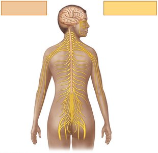
Functional Divisions of the PNS
Sensory (Afferent) Division: Transmits impulses from sensory receptors to the CNS. Includes:
Somatic sensory fibers: Convey impulses from skin, skeletal muscles, and joints.
Visceral sensory fibers: Convey impulses from visceral organs.
Motor (Efferent) Division: Transmits impulses from the CNS to effector organs (muscles and glands). Subdivided into:
Somatic Nervous System: Voluntary control of skeletal muscles.
Autonomic Nervous System (ANS): Involuntary control of smooth muscle, cardiac muscle, and glands. Includes:
Sympathetic Division: Mobilizes body systems during activity.
Parasympathetic Division: Conserves energy and promotes housekeeping functions during rest.
Histology of Nervous Tissue
Principal Cell Types
Nervous tissue consists of two main cell types:
Neuroglia (Glial Cells): Support, protect, and insulate neurons.
Neurons (Nerve Cells): Excitable cells that transmit electrical signals.
Neuroglia of the CNS
Astrocytes: Most abundant, versatile, and highly branched glial cells. They support neurons, regulate the chemical environment, guide neuron migration, and participate in information processing.
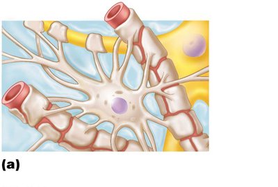
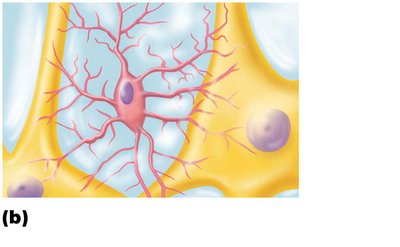
Microglial Cells: Small, ovoid cells with thorny processes. They monitor neuron health, migrate toward injured neurons, and can transform into phagocytes to remove debris and pathogens.
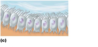
Ependymal Cells: Line the central cavities of the brain and spinal cord, often ciliated to help circulate cerebrospinal fluid (CSF), and form a barrier between CSF and tissue fluid.
Oligodendrocytes: Branched cells that form insulating myelin sheaths around CNS nerve fibers.
Neuroglia of the PNS
Satellite Cells: Surround neuron cell bodies in the PNS, functioning similarly to astrocytes in the CNS.
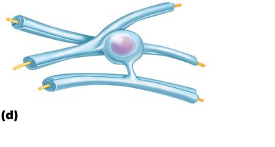
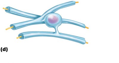
Schwann Cells (Neurolemmocytes): Surround all peripheral nerve fibers, form myelin sheaths in thicker fibers, and are vital for regeneration of damaged peripheral nerve fibers.

Neurons: Structure and Function
General Characteristics
Longevity: Neurons can last a lifetime.
Amitotic: Most do not divide after development (few exceptions).
High Metabolic Rate: Require continuous oxygen and glucose supply.
Basic Structure: All neurons have a cell body and one or more processes (dendrites and axons).
Neuron Cell Body (Perikaryon or Soma)
Biosynthetic Center: Synthesizes proteins, membranes, and chemicals; contains rough ER (Nissl bodies).
Nucleus: Spherical with a prominent nucleolus.
Location: Most neuron cell bodies are in the CNS (nuclei); in the PNS, they are found in ganglia.
Neuron Processes
Dendrites: Short, branched processes that receive input and convey signals toward the cell body as graded potentials.
Axon: Each neuron has one axon, which generates and transmits nerve impulses away from the cell body. Axons may branch and end in axon terminals, where neurotransmitters are released.

Axonal Transport: Movement of materials along the axon can be anterograde (away from cell body) or retrograde (toward cell body).
Myelin Sheath
Composition: Whitish, protein-lipid substance that insulates axons and increases the speed of impulse transmission.
Myelinated Fibers: Have segmented sheaths; conduct impulses rapidly.
Nonmyelinated Fibers: Lack myelin; conduct impulses more slowly.
Myelination in the PNS
Formed by Schwann cells wrapping around axons in a jelly roll fashion.
Myelin sheath gaps (nodes of Ranvier) are sites where axon collaterals can emerge.
Nonmyelinated fibers are thin and not wrapped in myelin but are surrounded by Schwann cells.

Myelination in the CNS
Formed by oligodendrocyte processes; each cell can myelinate multiple axons.
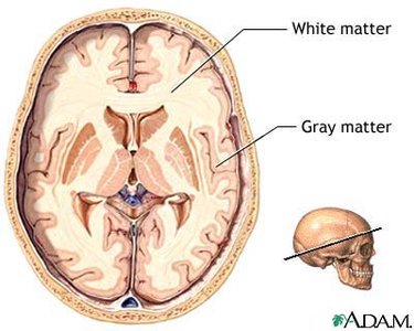
White matter consists of myelinated fibers; gray matter consists mostly of neuron cell bodies and non-myelinated fibers.
Classification of Neurons
Structural Classification
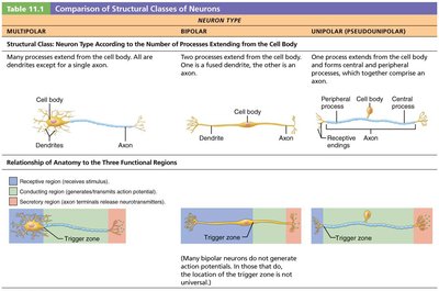
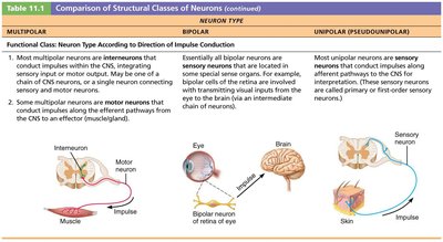
Multipolar Neurons: Three or more processes (one axon, others dendrites); most common in CNS.
Bipolar Neurons: Two processes (one axon, one dendrite); rare, found in retina and olfactory mucosa.
Unipolar (Pseudounipolar) Neurons: One T-like process (two axons); found mainly in PNS sensory ganglia.
Neuron Type | Processes | Location |
|---|---|---|
Multipolar | 1 axon, many dendrites | CNS (most abundant) |
Bipolar | 1 axon, 1 dendrite | Retina, olfactory mucosa |
Unipolar | 1 T-like process (2 axons) | PNS sensory ganglia |
Functional Classification
Sensory (Afferent) Neurons: Transmit impulses from sensory receptors toward the CNS; almost all are unipolar, with cell bodies in PNS ganglia.
Motor (Efferent) Neurons: Carry impulses from the CNS to effectors; multipolar, with most cell bodies in the CNS.
Interneurons (Association Neurons): Lie between sensory and motor neurons, shuttle signals through CNS pathways, and comprise 99% of all neurons.
Additional info: The nervous system's organization and cellular composition are foundational for understanding higher-level processes such as reflexes, sensation, and voluntary movement, which are covered in subsequent chapters.
