 Back
BackFundamentals of the Nervous System: Structure and Function
Study Guide - Smart Notes
 Tailored notes based on your materials, expanded with key definitions, examples, and context.
Nervous System Overview
The nervous system is essential for detecting and responding to changes in both the internal and external environments of an organism. As vertebrates evolved, specialized cells and tissues developed to rapidly process information and coordinate responses. The nervous system is divided into the central nervous system (CNS), peripheral nervous system (PNS), and sense organs. This guide focuses on the cellular and tissue organization of the nervous system, emphasizing the structure and function of neurons and supporting cells.
Nervous System Tissues
Basic Neuron and Receptors
Neurons are the primary cells responsible for receiving, integrating, and transmitting information. Receptors, which may be free nerve endings or specialized cells, detect environmental changes and convert them into electrical signals.
Free nerve endings: Primitive receptors directly impacted by the environment.
Specialized receptor cells: Act as transducers, converting one form of energy to another.
Resting potential: All neurons maintain an electrical potential across their membrane due to ion distribution.
Phasic receptors: Respond to changes in stimulus intensity; generate graded potentials proportional to stimulus magnitude.
Tonic receptors: Continuously active; fire only when a threshold is reached.
All-or-nothing receptors: Fire only under specific conditions; stimulus magnitude is encoded by frequency or pattern.
Neural Tissue Cell Types
Neural tissue consists of two main cell types: neurons and supporting cells (neuroglia or glial cells). Glial cells provide structural and metabolic support, insulation, and protection for neurons.
CNS Neuroglia
Astrocytes: Regulate ionic composition, maintain extracellular fluid balance, and support nutrition by contacting capillaries and neurons.
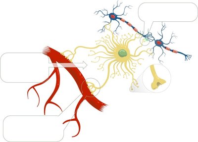
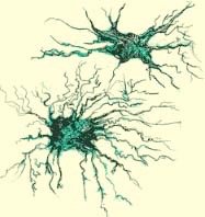
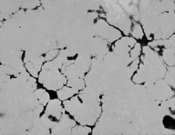
Microglia: Small, oval-shaped cells with thorny processes; act as macrophages to remove debris and pathogens.
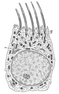
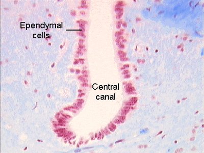
Ependymal cells: Line the central cavities of the brain and spinal cord; form a barrier between cerebrospinal fluid and CNS tissue fluid; often ciliated to move fluids.
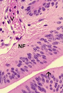
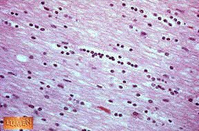
Oligodendrocytes: Produce myelin sheaths in the CNS; myelinated axons form white matter, while unmyelinated regions and cell bodies form gray matter.
PNS Neuroglia
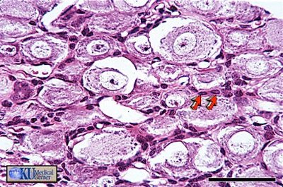
Satellite cells: Regulate the external chemical environment around PNS neurons.
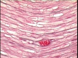
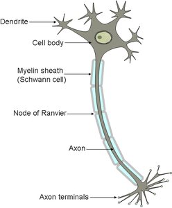
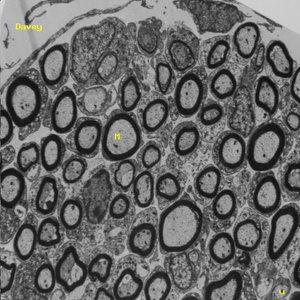
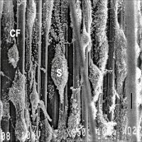
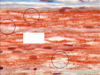
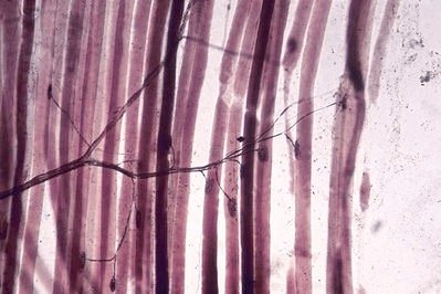
Schwann cells: Form myelin sheaths around PNS axons; essential for nerve fiber regeneration; myelin sheaths are interrupted by nodes of Ranvier.
Structural Organization: CNS vs. PNS
Structure | PNS Name | CNS Name | Glial Cells Responsible |
|---|---|---|---|
Aggregation of cell bodies | Ganglion | Nucleus or cortex (gray matter) | Astrocytes, microglia, ependymal cells |
Bundle of myelinated axons | Nerve | Tract | Schwann cells (PNS), Oligodendrocytes (CNS) |
Neurons: Structure and Function
Neurons are the primary functional units of the nervous system. Despite their diversity, all neurons share four main structural features:
Cell body (soma): Contains the nucleus and metabolic machinery; site of protein and neurotransmitter synthesis.
Dendrites: Receive stimuli and initiate graded potentials; increase surface area for synaptic input.
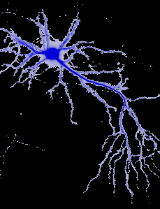
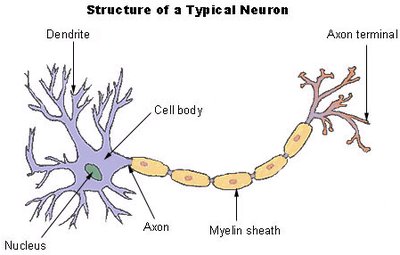
Axon: Long, slender process that conducts nerve impulses away from the cell body; may be myelinated or unmyelinated.
Terminal arborizations (axon terminals): Branching ends of the axon that form synapses with other neurons or effectors; contain synaptic vesicles with neurotransmitters.
Types of Neurons
Multipolar motor neurons: Cell body near dendrites, long axon extends outward.
Bipolar neurons: Cell body located between two processes (axon and dendrite).
Pseudounipolar sensory neurons: Cell body off to one side of the axon.
Neuronal Stimulation and Signal Transmission
Neurons communicate via electrical and chemical signals. The process begins with depolarization, which, if reaching threshold, triggers an action potential.
Depolarization: Opening of membrane channels in response to chemical, voltage, or mechanical stimuli; must reach threshold for action potential generation.
Graded potentials: Local changes in membrane potential; magnitude varies with stimulus strength and decreases with distance.
Action potentials: All-or-nothing electrical impulses that propagate along the axon; intensity encoded by frequency, not amplitude.
Conduction speed: Determined by axon diameter and degree of myelination.
Synapses
Synapses are specialized junctions where neurons communicate with other neurons or effectors. They can be electrical or chemical:
Electrical synapses: Allow direct, rapid transmission of signals via gap junctions; found throughout the CNS.
Chemical synapses: Use neurotransmitters released from synaptic vesicles to transmit signals across the synaptic cleft to postsynaptic receptors.
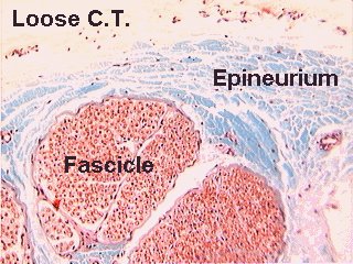
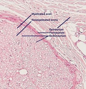
Types of synaptic connections include axodendritic, axosomatic, axoaxonic, dendrodendritic, and dendrosomatic. Neurons are classified as presynaptic (sending) or postsynaptic (receiving).
Key Equations
Nernst Equation (for equilibrium potential):
Ohm's Law (for membrane current):
Additional info: The notes above expand on the original content by providing definitions, examples, and academic context for each cell type and process. Images are included only where they directly reinforce the explanation of the adjacent paragraph.
