Back
BackFundamentals of the Nervous System: Structure and Function
Study Guide - Smart Notes
 Tailored notes based on your materials, expanded with key definitions, examples, and context.
General Functions of the Nervous System
Overview of Nervous System Functions
The nervous system is responsible for monitoring internal and external environments, integrating sensory information, and coordinating voluntary and involuntary responses. It also supports higher-order processes such as thought, emotion, and learning.
Monitoring Stimuli: Detects changes inside and outside the body.
Integration: Processes and interprets sensory input, integrating bodily functions.
Control of Effectors: Directs voluntary (skeletal muscle) and involuntary (smooth muscle, cardiac muscle, glands) responses.
Higher Functions: Supports conscious thought, perception, emotions, learning, and behavior.
Basic Function of the Nervous System
Three Main Functions
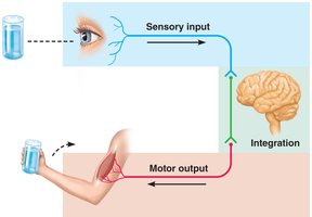
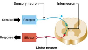
The nervous system operates through three fundamental steps: sensory input, integration, and motor output.
Sensory Input: Sensory receptors detect changes and send information to the central nervous system (CNS).
Integration: The CNS processes and interprets sensory input, making decisions about appropriate responses.
Motor Output: The CNS sends commands to effectors (muscles or glands) to elicit a response.
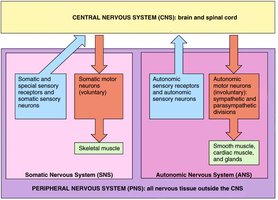
Anatomical Divisions of the Nervous System
Central and Peripheral Nervous Systems
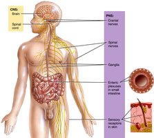
The nervous system is divided into the central nervous system (CNS) and the peripheral nervous system (PNS), each with distinct structures and functions.
Central Nervous System (CNS): Composed of the brain and spinal cord; responsible for processing and integrating information.
Peripheral Nervous System (PNS): Consists of cranial and spinal nerves; transmits sensory information to the CNS and carries motor commands to effectors.
Functional Divisions and Subdivisions of the Nervous System
Somatic and Autonomic Nervous Systems
The PNS is functionally divided into afferent (sensory) and efferent (motor) divisions, with further subdivisions based on the type of effectors controlled.
Afferent (Sensory) Division: Conducts impulses from sensory receptors to the CNS.
Efferent (Motor) Division: Conducts impulses from the CNS to effectors; subdivided into:
Somatic Nervous System (SNS): Controls voluntary movements of skeletal muscles.
Autonomic Nervous System (ANS): Regulates involuntary functions of smooth muscle, cardiac muscle, and glands.
Sympathetic Division: Prepares the body for 'fight or flight' responses.
Parasympathetic Division: Promotes 'rest and digest' activities.
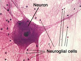
Nervous System Histology
Cells of the Nervous System
The nervous system is composed of two main cell types: neurons and neuroglia.
Neurons: Functional cells that conduct nerve impulses and form synapses with other neurons or effectors.
Neuroglia (Glial Cells): Supportive cells that maintain the health and function of neurons.
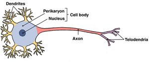
General Anatomy of a Neuron
Neuron Structure
Neurons have specialized structures for receiving, processing, and transmitting information.
Cell Body (Soma): Contains the nucleus and perikaryon (cytoplasm with organelles such as Nissl substance).
Dendrites: Branching extensions that receive signals from other neurons.
Axon: Long process that transmits action potentials away from the cell body; may branch into collateral axons and ends in telodendria with synaptic terminals.
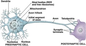
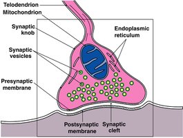
Synapse
Structure and Function of Synapses
A synapse is the junction between two neurons or between a neuron and an effector cell. It enables the transmission of nerve impulses via neurotransmitters stored in synaptic vesicles.
Presynaptic Terminal: Contains synaptic vesicles filled with neurotransmitter.
Synaptic Cleft: Small gap between presynaptic and postsynaptic membranes.
Postsynaptic Membrane: Receives the neurotransmitter and initiates a response in the next cell.
Axoplasmic Transport
Movement of Materials in Neurons
Axoplasmic transport is the process by which materials are moved between the neuron cell body and synaptic terminals.
Anterograde Flow: Movement from the cell body to the synaptic terminal; delivers neurotransmitters, enzymes, and lysosomes.
Retrograde Flow: Movement from the synaptic terminal toward the cell body; removes debris and can be exploited by pathogens.
Structural Classification of Neurons
Types of Neurons by Structure
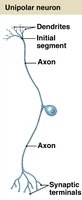
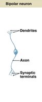
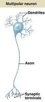
Neurons are classified based on the number of processes extending from the cell body.
Type | Description | Location/Function |
|---|---|---|
Unipolar | Single process (axon) attached to cell body | Most sensory (afferent) neurons |
Bipolar | Two processes (dendrite and axon) | Special sense organs (e.g., eyes) |
Multipolar | Multiple dendrites, one axon | All motor (efferent) neurons and most interneurons |
Functional Classification of Neurons
Types of Neurons by Function
Neurons are also classified by the direction in which they conduct nerve impulses.
Sensory (Afferent) Neurons: Conduct impulses from sensory receptors to the CNS; typically unipolar or bipolar.
Motor (Efferent) Neurons: Conduct impulses from the CNS to effectors (muscles or glands); typically multipolar.
Interneurons (Association Neurons): Relay impulses between neurons; most abundant and primarily located in the CNS.
Organization of Neurons in the CNS and PNS
Gray Matter, White Matter, Nerves, and Ganglia
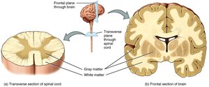
Neurons are organized into distinct structures in the CNS and PNS.
CNS: Neuron cell bodies form nuclei (gray matter); axons form tracts (white matter).
PNS: Neuron cell bodies form ganglia; axons are bundled into nerves.
Nerves
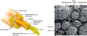
Structure of Peripheral Nerves
Nerves are bundles of axons in the PNS, containing both sensory and motor fibers, blood vessels, and connective tissue coverings.
Epineurium: Outermost connective tissue covering the entire nerve.
Perineurium: Surrounds bundles of axons (fascicles).
Endoneurium: Encloses individual axons and their Schwann cells.
Neuroglia (Glial Cells)
Types and Functions of Neuroglia
Neuroglia are supportive cells in the CNS and PNS, each with specialized roles.
CNS Neuroglia: Astrocytes, ependymal cells, oligodendrocytes, microglia
PNS Neuroglia: Schwann cells, satellite cells
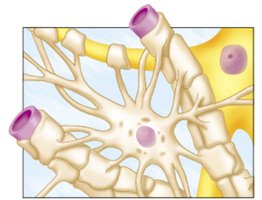
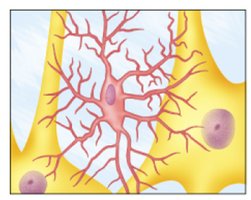
Astrocytes
Astrocytes anchor neurons to blood vessels, regulate the neuronal microenvironment, transport nutrients and wastes, and contribute to the blood-brain barrier.
Ependymal Cells
Ependymal cells line the ventricles of the brain and central canal of the spinal cord, producing and circulating cerebrospinal fluid (CSF). They also help form the blood-brain barrier.
Oligodendrocytes
Oligodendrocytes produce the myelin sheath in the CNS, which insulates axons and increases the speed of nerve impulse conduction.
Microglia
Microglia are phagocytic cells that defend the CNS against pathogens and remove debris.
Satellite and Schwann Cells (PNS)
Satellite cells surround neuron cell bodies in ganglia, maintaining their microenvironment. Schwann cells produce the myelin sheath in the PNS, enabling rapid saltatory conduction and aiding in axonal regeneration.
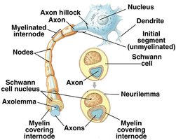
Myelinated Motor Neuron
Structure and Function of Myelinated Neurons
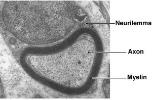
Myelinated neurons have axons wrapped in a myelin sheath, which increases the speed of action potential conduction and provides insulation.
Nodes of Ranvier: Gaps in the myelin sheath where action potentials are regenerated, enabling saltatory conduction.
Schwann Cells: Form the myelin sheath in the PNS; each cell myelinates a segment of one axon.
Neurilemma: The outermost layer of the Schwann cell, important for axonal regeneration.