 Back
BackFundamentals of the Nervous System: Structured Study Notes
Study Guide - Smart Notes
 Tailored notes based on your materials, expanded with key definitions, examples, and context.
Nervous System Fundamentals
Functions of the Nervous System
The nervous system is responsible for coordinating the body's activities through three primary functions: sensory input, integration, and motor output.
Sensory Input: The detection of internal and external stimuli by sensory receptors.
Integration: Processing and interpretation of sensory input to determine an appropriate response.
Motor Output: Activation of effector organs (muscles and glands) to produce a response.
Structural and Functional Divisions of the Nervous System
The nervous system is organized into central and peripheral divisions, each with distinct structural and functional roles.
Central Nervous System (CNS): Composed of the brain and spinal cord; responsible for integration and command.
Peripheral Nervous System (PNS): Consists of cranial and spinal nerves; connects the CNS to the rest of the body.
Afferent (Sensory) Division: Transmits sensory information to the CNS.
Efferent (Motor) Division: Carries motor commands from the CNS to effectors.
Somatic Nervous System: Controls voluntary movements of skeletal muscles.
Autonomic Nervous System: Regulates involuntary functions; subdivided into sympathetic and parasympathetic systems.
Neurons and Neuroglia
Types of Neuroglia and Their Functions
Neuroglia are supportive cells in the nervous system, essential for neuron function and health.
CNS Neuroglia:
Astrocytes: Support neurons, regulate ion balance, and maintain the blood-brain barrier.
Microglia: Act as immune cells, removing debris and pathogens.
Ependymal Cells: Line ventricles and produce cerebrospinal fluid.
Oligodendrocytes: Form myelin sheaths in the CNS.
PNS Neuroglia:
Satellite Cells: Surround neuron cell bodies in ganglia, regulate environment.
Schwann Cells: Form myelin sheaths in the PNS.
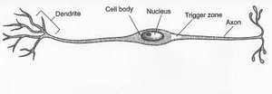
Neuron Structure and Function
Neurons are the fundamental units of the nervous system, specialized for transmitting electrical signals.
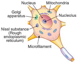
Cell Body (Soma): Contains the nucleus, nucleolus, mitochondria, Golgi apparatus, Nissl substance (rough endoplasmic reticulum), and microfilaments for structural support.
Dendrites: Receive incoming signals.
Axon: Conducts electrical impulses away from the cell body.
Myelin Sheath
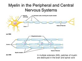
The myelin sheath is a protective covering that insulates axons, increasing the speed of nerve impulse transmission.
Formation in the PNS: Schwann cells wrap around axons, forming the myelin sheath.
Formation in the CNS: Oligodendrocytes extend processes to multiple axons, creating myelin segments.
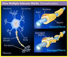
Role in Multiple Sclerosis: In MS, myelin is damaged, leading to impaired nerve conduction.
Classification of Neurons
Neurons are classified by structure and function.
Structural Classification:
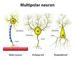
Multipolar Neurons: Many dendrites, one axon; most common in CNS.
Bipolar Neurons: One dendrite, one axon; found in sensory organs.
Unipolar Neurons: Single process; common in PNS sensory neurons.
Functional Classification:
Sensory (Afferent) Neurons: Transmit impulses to CNS.
Motor (Efferent) Neurons: Carry impulses from CNS to effectors.
Interneurons (Association Neurons): Connect sensory and motor neurons within CNS.
Nerve vs. Tract & Nucleus vs. Ganglion
Understanding terminology is essential for distinguishing structures in the nervous system.
Nerve: Bundle of axons in the PNS.
Tract: Bundle of axons in the CNS.
Nucleus: Cluster of neuron cell bodies in the CNS.
Ganglion: Cluster of neuron cell bodies in the PNS.
Connective Tissue Coverings
Nerves are protected by three layers of connective tissue:
Epineurium: Surrounds the entire nerve.
Perineurium: Encloses bundles of axons (fascicles).
Endoneurium: Surrounds individual axons.
Bioelectricity in Neurons
Principles of Electricity
Neurons use electrical signals to communicate. Key concepts include voltage, current, and resistance.
Voltage (V): Potential energy from separated charges; measured in volts (V) or millivolts (mV).
Current (I): Flow of electric charge; used to do work.
Resistance (R): Opposition to current flow; insulators have high resistance, conductors have low resistance.
Relationship:
Ohm's Law:
Membrane Ion Channels
Ion channels regulate the movement of ions across the neuronal membrane, crucial for electrical signaling.
Leaky Channels: Allow passive ion movement.
Gated Channels: Open in response to specific stimuli. Types include chemically-gated (ligand), voltage-gated, and mechanically-gated channels.
Resting Membrane Potential
The resting membrane potential is the voltage difference across the membrane when the neuron is not transmitting signals.
Typical values range from -40 mV to -90 mV.
Established by differences in ion composition and membrane permeability.
Maintained by the sodium-potassium pump ( out, in).
Graded Potentials
Graded potentials are short-lived, localized changes in membrane potential, essential for initiating action potentials.
Strength depends on stimulus intensity.
Dissipate with distance from origin.
Act as signals over short distances.
Action Potentials
Action potentials are the principal means by which neurons send long-distance signals.
Brief reversal of membrane potential (~100 mV).
All-or-none response: occurs fully or not at all.
Phases: depolarization, repolarization, hyperpolarization.
Generation and Propagation of Action Potentials
Resting State: All gated channels closed; only K+ leakage channels open.
Depolarization: Voltage-gated Na+ channels open; Na+ influx causes membrane to become less negative.
Threshold: Minimum change (~15–20 mV) required to trigger action potential.
Repolarization: Na+ channels close; K+ channels open, K+ exits cell.
After-hyperpolarization: Excess K+ outflow overshoots resting potential.
Na+/K+ Pump: Restores original ion concentrations.
Propagation
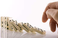
Action potentials propagate along axons like falling dominos, triggering adjacent segments.
Refractory Periods
Absolute Refractory Period: No new action potential can be generated.
Relative Refractory Period: Action potential possible with stronger stimulus.
Conduction Velocity
Axon Diameter: Larger diameter = faster conduction.
Degree of Myelination:
Continuous Conduction: Unmyelinated fibers; slower.
Saltatory Conduction: Myelinated fibers; faster, jumps between nodes of Ranvier.
Synapses
Definition and Structure
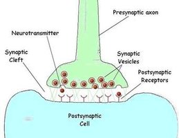
A synapse is a junction mediating information transfer between neurons or from neuron to effector cell.
Presynaptic Membrane: Releases neurotransmitter.
Synaptic Cleft: Gap between neurons.
Postsynaptic Membrane: Receives neurotransmitter.
Chemical vs. Electrical Synapses
Chemical Synapses: Use neurotransmitters; most common.
Electrical Synapses: Use gap junctions; direct electrical flow.
Postsynaptic Potentials
Excitatory Postsynaptic Potentials (EPSPs): Depolarize membrane, increase likelihood of action potential.
Inhibitory Postsynaptic Potentials (IPSPs): Hyperpolarize membrane, decrease likelihood of action potential.
Summation:
Temporal Summation: Rapid succession of EPSPs.
Spatial Summation: Multiple EPSPs from different locations.
Facilitation: Substance brings neuron closer to threshold.
Neurotransmitters
Neurotransmitters are chemical messengers released at synapses, classified by structure and function.
Acetylcholine
Biogenic Amines: Norepinephrine, dopamine, serotonin, histamine
Amino Acids: GABA, glutamate
Peptides: Endorphins, enkephalin, dynorphin, substance P
Purines: ATP, adenosine
Gases and Lipids: Nitric oxide, carbon monoxide
Neurotransmitters can be excitatory or inhibitory, and their actions can be modified by various substances.
Neurotransmitter | Class | Function |
|---|---|---|
Acetylcholine | Choline derivative | Excitatory at neuromuscular junctions |
Norepinephrine | Biogenic amine | Excitatory or inhibitory |
GABA | Amino acid | Inhibitory |
Glutamate | Amino acid | Excitatory |
Endorphins | Peptide | Inhibitory, pain relief |
ATP | Purine | Excitatory or inhibitory |
Nitric oxide | Gas | Excitatory |
Summary Table: Nervous System Organization
Division | Components | Function |
|---|---|---|
CNS | Brain, Spinal Cord | Integration, command |
PNS | Cranial & Spinal Nerves | Connects CNS to body |
Somatic | Skeletal Muscles | Voluntary movement |
Autonomic | Smooth/Cardiac Muscles, Glands | Involuntary functions |
Sympathetic | Various organs | Fight or flight |
Parasympathetic | Various organs | Rest and digest |
Additional info: Academic context and explanations have been expanded for clarity and completeness. Images included are directly relevant to the adjacent paragraphs and reinforce key concepts.
