 Back
BackGuided Study for Chapter 3: The Cell (Anatomy & Physiology)
Study Guide - Smart Notes
 Tailored notes based on your materials, expanded with key definitions, examples, and context.
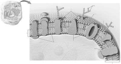
Q1. Identify and color-code each component of the plasma membrane in Figure 3.1. Then, list the main function(s) of each component.
Background
Topic: Structure and Function of the Plasma Membrane
This question tests your understanding of the plasma membrane's structure, including its major components (lipids, proteins, carbohydrates) and their functions according to the fluid mosaic model.
Key Terms and Formulas:
Phospholipid bilayer: Double layer of phospholipids forming the basic structure of the membrane.
Integral proteins: Proteins embedded within the membrane, often spanning it.
Peripheral proteins: Proteins attached to the membrane surface.
Carbohydrate chains: Attached to proteins or lipids, involved in cell recognition.
Cholesterol: Stabilizes membrane fluidity.
Step-by-Step Guidance
Examine Figure 3.1 and identify the phospholipid bilayer, noting its amphipathic nature (hydrophilic heads and hydrophobic tails).
Locate and distinguish between integral and peripheral proteins in the membrane. Consider their placement and possible functions (e.g., transport, signaling).
Identify carbohydrate chains attached to proteins (glycoproteins) or lipids (glycolipids) and think about their role in cell recognition and signaling.
Find cholesterol molecules interspersed within the bilayer and consider how they affect membrane fluidity and stability.
For each component, list its main function(s) based on your textbook or glossary definitions.
Try solving on your own before revealing the answer!
Final Answer:
The plasma membrane consists of:
Phospholipid bilayer: Provides a semi-permeable barrier, allowing selective passage of substances.
Integral proteins: Function as channels, carriers, or receptors for transport and communication.
Peripheral proteins: Support membrane structure and participate in signaling.
Carbohydrate chains: Involved in cell recognition and signaling.
Cholesterol: Maintains membrane fluidity and stability.
Each component plays a critical role in maintaining the cell's integrity and facilitating communication and transport.
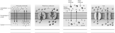
Q2. Identify the types of passive transport in the illustrations in Figure 3.2.
Background
Topic: Passive Transport Across the Plasma Membrane
This question tests your ability to recognize and differentiate between types of passive transport: simple diffusion, facilitated diffusion, and osmosis.
Key Terms and Formulas:
Simple diffusion: Movement of nonpolar molecules directly through the membrane.
Facilitated diffusion: Movement of polar molecules via protein channels or carriers.
Osmosis: Movement of water across the membrane.
Tonicity: Describes the effect of solution concentration on cell volume.
Step-by-Step Guidance
Look at each panel in Figure 3.2 and note the direction and type of molecules moving across the membrane.
Identify which panels show nonpolar molecules moving directly through the bilayer (simple diffusion).
Identify panels where polar molecules move through protein channels (facilitated diffusion).
Find the panel showing water movement (osmosis) and note the direction relative to solute concentration.
Label each type of passive transport under the corresponding illustration.
Try solving on your own before revealing the answer!
Final Answer:
First panel: Simple diffusion (nonpolar molecules)
Second panel: Facilitated diffusion (polar molecules)
Third panel: Osmosis (water movement)
Fourth panel: Ion channel (facilitated diffusion for ions)
Each type of passive transport allows substances to move down their concentration gradient without energy input.
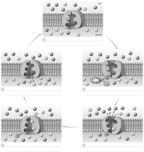
Q3. Sequence the Events: Stages of the Na+/K+ Pump in Figure 3.3.
Background
Topic: Primary Active Transport (Na+/K+ Pump)
This question tests your understanding of the steps involved in the sodium-potassium pump, a primary active transport mechanism that maintains cellular ion gradients.
Key Terms and Formulas:
Na+/K+ pump: Transports 3 Na+ out and 2 K+ into the cell using ATP.
ATP: Provides energy for active transport.
Concentration gradient: Difference in ion concentration across the membrane.
Step-by-Step Guidance
Observe Figure 3.3 and identify the initial binding of Na+ ions to the pump on the cytoplasmic side.
Note the phosphorylation of the pump by ATP, causing a conformational change.
Follow the release of Na+ ions to the extracellular side and the binding of K+ ions.
Track the dephosphorylation of the pump and the return of K+ ions to the cytoplasm.
Sequence the steps in your own words, color-coding Na+ and K+ as instructed.
Try solving on your own before revealing the answer!
Final Answer:
Three Na+ ions bind to the pump inside the cell.
ATP phosphorylates the pump, causing it to change shape and release Na+ outside.
Two K+ ions bind to the pump from outside.
The pump is dephosphorylated, returning to its original shape and releasing K+ inside.
This cycle maintains the electrochemical gradient essential for cell function.
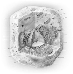
Q4. Practice Labeling: Parts of the Cell in Figure 3.4. Then, list the main function(s) of each component.
Background
Topic: Cell Structure and Organelles
This question tests your ability to identify major cell organelles and understand their functions within a generalized cell.
Key Terms and Formulas:
Nucleus: Stores genetic material and controls cell activities.
Mitochondria: Site of ATP production.
Ribosomes: Protein synthesis.
Endoplasmic reticulum (ER): Rough ER synthesizes proteins; smooth ER synthesizes lipids.
Golgi apparatus: Modifies, sorts, and packages proteins.
Lysosomes: Digest cellular waste.
Step-by-Step Guidance
Examine Figure 3.4 and identify each labeled organelle.
Recall the main function of each organelle based on your glossary or textbook.
Label each part and write its function next to it.
Consider how these organelles interact to maintain cell function.
Try solving on your own before revealing the answer!
Final Answer:
Nucleus: Contains DNA, directs cell activities.
Mitochondria: Produces ATP via cellular respiration.
Ribosomes: Synthesizes proteins.
Rough ER: Protein synthesis and processing.
Smooth ER: Lipid synthesis and detoxification.
Golgi apparatus: Modifies and packages proteins.
Lysosomes: Digests waste and cellular debris.
Each organelle plays a specific role in cell metabolism and maintenance.
Q5. Sequence the Events: Steps of Protein Synthesis in Figure 3.5.
Background
Topic: Protein Synthesis (Transcription and Translation)
This question tests your understanding of the steps involved in synthesizing proteins from DNA, including transcription and translation.
Key Terms and Formulas:
Transcription: DNA is copied into mRNA in the nucleus.
Translation: mRNA is decoded by ribosomes to build a polypeptide.
rRNA, mRNA, tRNA: Types of RNA involved in protein synthesis.
Codon: Three-base sequence on mRNA specifying an amino acid.
Step-by-Step Guidance
Review Figure 3.5 and identify the first step: transcription of DNA to mRNA in the nucleus.
Follow the export of mRNA from the nucleus to the cytoplasm.
Observe the binding of mRNA to ribosomes and the initiation of translation.
Track the role of tRNA in bringing amino acids to the ribosome, matching anticodons to mRNA codons.
Sequence the steps in your own words, describing the process up to polypeptide formation.
Try solving on your own before revealing the answer!
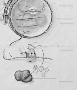
Final Answer:
Transcription: DNA is transcribed to mRNA in the nucleus.
mRNA exits the nucleus and binds to a ribosome.
Translation: tRNA brings amino acids to the ribosome, matching anticodons to mRNA codons.
A polypeptide chain is synthesized, forming a protein.
Protein synthesis is essential for cell function and structure.
Q6. Identify the phases of the cell cycle in Figure 3.6. Then, describe what happens during each phase.
Background
Topic: Cell Cycle and Division
This question tests your understanding of the stages of the cell cycle, including interphase, mitosis, and cytokinesis.
Key Terms and Formulas:
Cell cycle: Series of events from cell formation to division.
Interphase: G1 (growth), S (DNA synthesis), G2 (preparation for division).
Mitosis: Division of the nucleus.
Cytokinesis: Division of the cytoplasm.
Step-by-Step Guidance
Examine Figure 3.6 and identify each labeled phase: G1, S, G2, M, and G0.
Describe what happens during G1 (cell growth), S (DNA replication), and G2 (preparation for division).
Explain the events of mitosis (nuclear division) and cytokinesis (cytoplasmic division).
Note the significance of each phase for cell function and survival.
Try solving on your own before revealing the answer!

Final Answer:
G1 phase: Cell grows and prepares for DNA replication.
S phase: DNA is replicated.
G2 phase: Cell prepares for division.
M phase: Mitosis (nuclear division) and cytokinesis (cytoplasmic division).
G0 phase: Resting phase for non-dividing cells.
Each phase is essential for proper cell growth, replication, and division.
