 Back
BackHigher Cortical Functions and Disorders of the Nervous System
Study Guide - Smart Notes
 Tailored notes based on your materials, expanded with key definitions, examples, and context.
Higher Cortical Functions
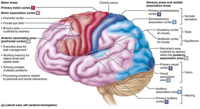
Cerebral Cortex: Structure and Function
The cerebral cortex is the executive suite of the brain, responsible for conscious mind activities such as awareness, sensory perception, voluntary motor initiation, communication, memory storage, and understanding. It is a thin (2–4 mm) superficial layer of gray matter, composed of neuron cell bodies, dendrites, glial cells, and blood vessels, but no axons. The cortex accounts for about 40% of the total brain mass.
Functional Areas: The cortex contains three types of functional areas:
Motor areas: Control voluntary movement
Sensory areas: Provide conscious awareness of sensation
Association areas: Integrate diverse information
Contralateral Control: Each hemisphere is concerned with the opposite side of the body.
Lateralization: Specialization of cortical function can occur in only one hemisphere.
Integration: Conscious behavior involves the entire cortex in some way.
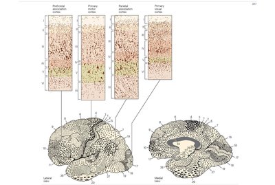
Example: The primary visual cortex has a prominent internal granular cell layer (layer IV), while the primary motor cortex has a prominent output layer (layer V). These differences are the basis for Brodmann's cytoarchitectonic regions.
Techniques to Study Brain Function
fMRI (Functional Magnetic Resonance Imaging): Measures brain activity by detecting changes in blood flow.
PET (Positron Emission Tomography): Uses radioactive tracers to visualize active brain regions.
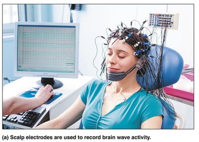
EEG (Electroencephalogram): Records electrical activity of the brain via scalp electrodes.
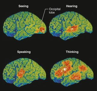
Example: fMRI and PET scans show that specific motor and sensory functions are located in discrete cortical areas called domains, while higher functions are distributed over many areas.
Diagnostic Procedures for CNS Dysfunction
Reflex Tests: Simple tests like the knee-jerk reflex can indicate CNS dysfunction.
CT and MRI: Identify tumors, lesions, plaques, or infarcts.
PET Scans: Localize brain lesions that generate seizures.
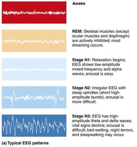
Brain Wave Patterns and the EEG
EEG and Brain Waves
The EEG records patterns of neuronal electrical activity generated by synaptic activity in the cortex. Each person's brain waves are unique and can change with age, sensory stimuli, brain disease, and chemical state of the body. EEGs are used to diagnose epilepsy, sleep disorders, and to determine brain death.
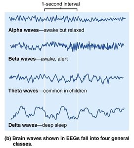
Wave Frequency: Measured in Hertz (Hz), or cycles per second.
Four Classes of Brain Waves:
Alpha waves (8–13 Hz): Regular, rhythmic, low-amplitude; indicate an idling brain.
Beta waves (14–30 Hz): Rhythmic, less regular; occur when mentally alert.
Theta waves (4–7 Hz): More irregular; common in children, uncommon in awake adults.
Delta waves (<4 Hz): High-amplitude; seen in deep sleep or brain damage in awake adults.
Higher Mental Functions
Language
The language implementation system involves the association cortex of the left hemisphere. Main areas include:
Broca’s area: Involved in speech production. Lesions cause inability to speak but comprehension remains intact.
Wernicke’s area: Involved in understanding spoken and written words. Lesions cause fluent but nonsensical speech.
Corresponding areas on the right side are involved with nonverbal language components.
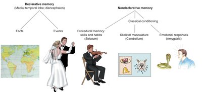
Learning and Memory
Memory is the storage and retrieval of information. There are several types:
Declarative (fact) memory: Names, faces, words, dates.
Procedural (skills) memory: Skills like playing piano.
Motor memory: Motor skills such as riding a bike.
Emotional memory: Experiences linked to emotions (e.g., fear response to a snake).
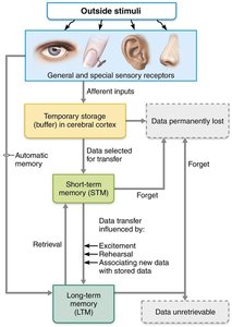
Declarative memory storage occurs in two stages:
Short-term memory (STM): Temporary holding, limited to 7–8 items.
Long-term memory (LTM): Limitless capacity.
Factors affecting transfer from STM to LTM include emotional state, rehearsal, association, and automatic memory. Memory consolidation involves the hippocampus, temporal cortical areas, thalamus, and prefrontal cortex.
Clinical Aspects of Memory
Anterograde amnesia: Inability to form new memories; old memories remain intact.
Retrograde amnesia: Loss of memories formed in the distant past.
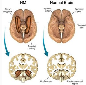
Example: The famous patient H.M. had bilateral removal of the hippocampus, resulting in profound anterograde amnesia.
Consciousness
Levels and Disorders of Consciousness
Consciousness involves perception of sensation, voluntary movement, and higher mental processing. It is clinically graded as:
Alertness
Drowsiness (lethargy)
Stupor
Coma
Loss of consciousness (except during sleep) signals impaired brain function. Fainting (syncope) is brief, while coma is prolonged unconsciousness. Brain death is an irreversible coma.
Epilepsy
Epileptic seizures are caused by abnormal electrical discharges in the brain. Types include:
Absence seizures: Brief, mild, often in children.
Tonic-clonic seizures: Severe, with convulsions and loss of consciousness.
Treatment includes anticonvulsant drugs and, in some cases, vagus nerve or deep brain stimulation.
Sleep and Sleep-Wake Cycles
Types and Stages of Sleep
Sleep is a state of partial unconsciousness. Two major types are defined by EEG patterns:
Non–rapid eye movement (non-REM) sleep: Stages N1, N2, N3 (slow-wave sleep).
Rapid eye movement (REM) sleep: Most dreaming occurs; temporary paralysis except for eye movements.

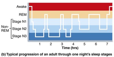
Sleep is regulated by the suprachiasmatic nucleus (biological clock) and preoptic nucleus (sleep-inducing center) of the hypothalamus. Orexins are hypothalamic chemicals that promote wakefulness.
Importance of Sleep
Consolidates new memories and discards unused ones.
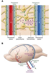
Restorative; non-REM sleep helps wash out brain waste products via cerebrospinal fluid.
Sleep deprivation leads to increased REM and slow-wave sleep in subsequent cycles.
Sleep Disorders
Narcolepsy: Abrupt lapses into sleep; associated with loss of orexins.
Insomnia: Chronic inability to obtain sufficient sleep; may be treated by blocking orexin action.
Diseases of the Nervous System
Brain Injuries and Disorders
Concussion: Temporary alteration in brain function.
Contusion: Permanent brain damage.
Hemorrhage: Subdural or subarachnoid bleeding increases intracranial pressure.
Cerebral edema: Swelling of the brain after injury.
Cerebrovascular Accidents (Strokes)
Ischemia: Loss of blood supply leads to brain tissue death, often due to arterial blockage.
Hemiplegia: Paralysis on one side of the body.
Transient Ischemic Attacks (TIAs): Temporary, reversible episodes of cerebral ischemia.
Degenerative Brain Disorders
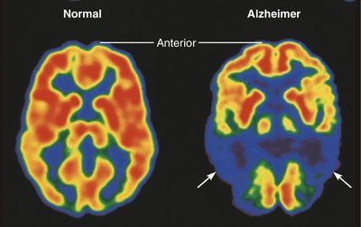
Alzheimer’s Disease (AD): Progressive degeneration causing dementia, memory loss, and brain shrinkage. Characterized by beta-amyloid plaques and neurofibrillary tangles (tau protein).
Parkinson’s Disease: Degeneration of dopamine-releasing neurons in the substantia nigra, leading to tremors and motor deficits. Treated with L-dopa, deep brain stimulation, or stem cell implants.
Huntington’s Disease: Fatal hereditary disorder due to accumulation of huntingtin protein, causing degeneration of basal nuclei and cortex, chorea, and mental deterioration.
