 Back
BackHistology and Function of Endocrine Glands and Blood Cells
Study Guide - Smart Notes
 Tailored notes based on your materials, expanded with key definitions, examples, and context.
Endocrine System: Glandular Histology and Function
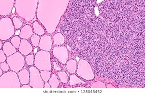
Thyroid Gland
The thyroid gland is a major endocrine organ located in the anterior neck, responsible for producing hormones that regulate metabolism and calcium balance. Its histological structure is characterized by spherical follicles filled with colloid, surrounded by follicular cells.
Follicular cells: Produce thyroid hormones (thyroxine T4 and triiodothyronine T3).
Colloid: Contains thyroglobulin, the precursor to thyroid hormones.
Parafollicular cells (C cells): Secrete calcitonin, which lowers blood calcium levels.
Function: Regulates metabolic rate, growth, and development.
Example: Hyperthyroidism results in increased metabolic rate, while hypothyroidism causes fatigue and weight gain.
Parathyroid Gland
The parathyroid glands are small, typically four in number, located on the posterior surface of the thyroid gland. They are essential for calcium homeostasis.
Chief cells: Secrete parathyroid hormone (PTH), which increases blood calcium levels.
Oxyphil cells: Function is less clear, but may play a role in PTH secretion.
Function: PTH stimulates osteoclasts, increases intestinal calcium absorption, and promotes kidney reabsorption of calcium.
Example: Parathyroid hormone deficiency leads to hypocalcemia and tetany.
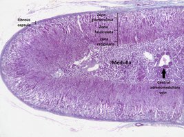
Adrenal Gland
The adrenal glands are paired organs located atop the kidneys, composed of two distinct regions: the cortex and medulla. Each region produces different hormones vital for stress response and homeostasis.
Adrenal cortex: Divided into three zones:
Zona glomerulosa: Produces mineralocorticoids (e.g., aldosterone).
Zona fasciculata: Produces glucocorticoids (e.g., cortisol).
Zona reticularis: Produces androgens.
Adrenal medulla: Produces catecholamines (epinephrine and norepinephrine).
Function: Regulates electrolyte balance, metabolism, and the fight-or-flight response.
Example: Addison's disease results from adrenal insufficiency; Cushing's syndrome from excess glucocorticoids.
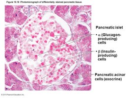
Pancreas and Islet of Langerhans
The pancreas is both an endocrine and exocrine organ. The Islets of Langerhans are clusters of endocrine cells within the pancreas that regulate blood glucose levels.
Alpha (α) cells: Secrete glucagon, which raises blood glucose.
Beta (β) cells: Secrete insulin, which lowers blood glucose.
Delta (δ) cells: Secrete somatostatin, which inhibits both insulin and glucagon secretion.
Pancreatic acinar cells: Exocrine cells that produce digestive enzymes.
Function: Maintains glucose homeostasis and aids digestion.
Example: Diabetes mellitus results from insufficient insulin production or action.
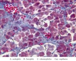
Anterior Pituitary (Adenohypophysis)
Cell Types and Hormones
The anterior pituitary is a key endocrine gland that produces several hormones regulating growth, metabolism, and reproduction. It contains distinct cell types identifiable by their staining properties.
Acidophils: Secrete growth hormone (GH) and prolactin.
Basophils: Secrete thyroid-stimulating hormone (TSH), adrenocorticotropic hormone (ACTH), follicle-stimulating hormone (FSH), and luteinizing hormone (LH).
Chromophobes: Cells with minimal staining, function less defined.
Function: Regulates growth, thyroid function, adrenal function, and reproductive processes.
Example: Pituitary adenomas can cause hormone imbalances, affecting multiple body systems.
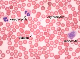
Blood Cells: Structure and Function
Types of Blood Cells
Blood is composed of various cell types, each with specialized functions in oxygen transport, immunity, and clotting.
Erythrocytes (red blood cells): Transport oxygen via hemoglobin.
Leukocytes (white blood cells): Include neutrophils, monocytes, lymphocytes, eosinophils, and basophils; function in immune defense.
Platelets: Cell fragments involved in blood clotting.
Function: Maintains oxygen delivery, immune protection, and hemostasis.
Example: Anemia results from reduced erythrocyte count; leukopenia from decreased white blood cells.
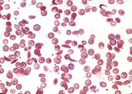
Blood Smear: Morphology
A blood smear is a laboratory technique used to examine the morphology of blood cells under a microscope. It helps diagnose various hematological disorders.
Normal morphology: Erythrocytes are biconcave, uniform in size; leukocytes are larger and less numerous.
Abnormal morphology: Variations in cell shape, size, or number can indicate disease (e.g., sickle cell anemia, leukemia).
Function: Provides diagnostic information for blood disorders.
Example: Sickle-shaped erythrocytes are characteristic of sickle cell anemia.
Summary Table: Endocrine Glands and Their Hormones
Gland | Main Hormones | Function |
|---|---|---|
Thyroid | T3, T4, Calcitonin | Metabolism, calcium regulation |
Parathyroid | PTH | Increases blood calcium |
Adrenal Cortex | Aldosterone, Cortisol, Androgens | Electrolyte balance, stress response |
Adrenal Medulla | Epinephrine, Norepinephrine | Fight-or-flight response |
Pancreas (Islets) | Insulin, Glucagon, Somatostatin | Glucose regulation |
Anterior Pituitary | GH, PRL, TSH, ACTH, FSH, LH | Growth, metabolism, reproduction |
Key Equations
Blood Glucose Regulation:
Calcium Regulation:
Additional info: Academic context was added to clarify gland functions, cell types, and clinical relevance for exam preparation.
