 Back
BackHistology: Structure and Function of Tissues
Study Guide - Smart Notes
 Tailored notes based on your materials, expanded with key definitions, examples, and context.
Histology: The Study of Tissues
Definition and Overview
Histology is the branch of anatomy that studies tissues, which are groups of cells with similar structure and function. There are four primary tissue types in the human body: epithelial, connective, muscle, and nervous tissue.
Epithelial Tissue: Forms protective boundaries and is involved in absorption, secretion, and sensation.
Connective Tissue: Provides support, protection, and structure; connects tissues together.
Muscle Tissue: Responsible for movement and essential bodily functions.
Nervous Tissue: Specialized for communication via electrical and chemical signals.
Epithelial Tissue
Functions of Epithelial Tissue
Epithelial tissue covers body surfaces, lines cavities, and forms glands. Its main functions include:
Protection: Shields underlying tissues from mechanical and thermal injury; produces keratin for added strength.
Immune Defense: Contains immune cells to defend against pathogens.
Secretion: Forms glands that produce substances such as sweat, oil, and hormones.
Transport: Acts as selectively permeable barriers for substance exchange.
Sensation: Contains nerve endings for detecting environmental changes.
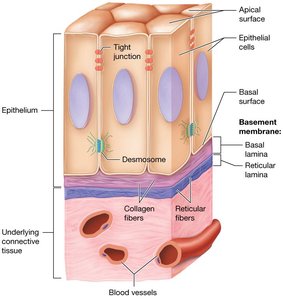
Structure of Epithelial Tissue
Epithelial cells are tightly packed and rest on a specialized basement membrane that separates them from underlying connective tissue. The basement membrane consists of:
Basal Lamina: ECM of epithelial tissue, rich in collagen fibers and ground substance.
Reticular Lamina: Produced by underlying connective tissue, contains reticular fibers.
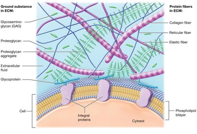
Extracellular Matrix (ECM) in Epithelial Tissue
The extracellular matrix (ECM) is composed of ground substance and protein fibers, providing structural and biochemical support to surrounding cells.
Ground Substance: Gel-like material containing water, ions, nutrients, and macromolecules such as glycosaminoglycans (GAGs), proteoglycans, and glycoproteins.
Protein Fibers: Collagen (strength), elastic (flexibility), and reticular fibers (supportive network).
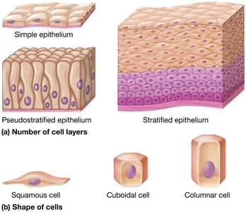
Classification of Epithelia
Epithelia are classified based on the number of cell layers and the shape of the cells:
Number of Layers:
Simple: One layer of cells
Stratified: Multiple layers
Pseudostratified: Appears multilayered but is a single layer
Cell Shape:
Squamous: Flattened
Cuboidal: Cube-shaped
Columnar: Tall and column-like
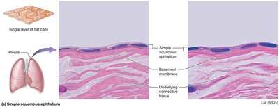
Types of Simple Epithelia
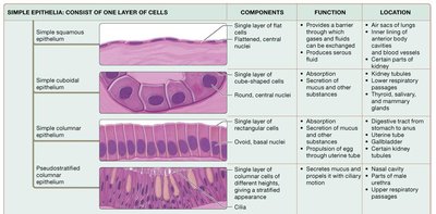
Simple Squamous Epithelium: Single layer of flat cells; allows diffusion and filtration. Found in alveoli, glomeruli, and blood vessels.
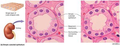
Simple Cuboidal Epithelium: Single layer of cube-shaped cells; functions in secretion and absorption. Found in kidney tubules and gland ducts.
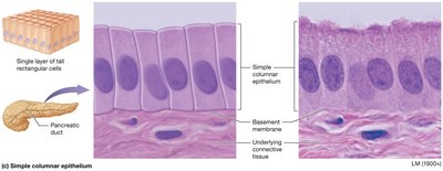
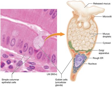
Simple Columnar Epithelium: Single layer of tall cells; may have microvilli or goblet cells. Functions in absorption and secretion. Found in the digestive tract and uterus.
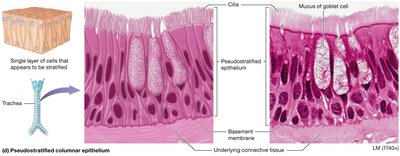
Pseudostratified Columnar Epithelium: Appears stratified but is a single layer; often ciliated with goblet cells. Functions in secretion and movement of mucus. Found in respiratory tract.
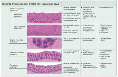
Types of Stratified Epithelia
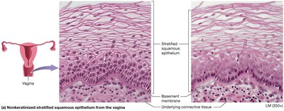
Stratified Squamous Epithelium: Multiple layers; protects underlying tissues. Keratinized type forms the skin, non-keratinized lines moist surfaces (mouth, vagina).
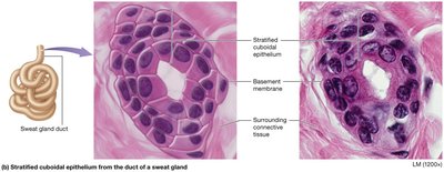
Stratified Cuboidal Epithelium: Two layers of cube-shaped cells; found in ducts of sweat glands.
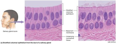
Stratified Columnar Epithelium: Multiple layers with columnar surface cells; rare, found in male urethra and some gland ducts.
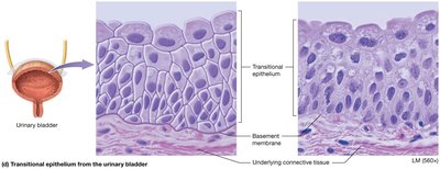
Transitional Epithelium: Multiple layers of cells that can change shape; allows stretching. Found in urinary bladder.
Summary Tables: Epithelial Tissue Types
Type | Components | Function | Location |
|---|---|---|---|
Simple squamous | Single layer of flat cells | Diffusion, filtration | Alveoli, blood vessels, serous membranes |
Simple cuboidal | Single layer of cube-shaped cells | Secretion, absorption | Kidney tubules, gland ducts |
Simple columnar | Single layer of tall cells | Absorption, secretion | Digestive tract, uterus |
Pseudostratified columnar | Single layer, appears stratified | Secretion, movement of mucus | Respiratory tract |
Stratified squamous | Multiple layers, flat surface cells | Protection | Skin, mouth, esophagus, vagina |
Stratified cuboidal | Two layers of cube-shaped cells | Protection | Sweat gland ducts |
Stratified columnar | Multiple layers, columnar surface cells | Protection, secretion | Male urethra, gland ducts |
Transitional | Multiple layers, variable shape | Stretching | Urinary bladder |
Glandular Epithelium
Types of Glands
Endocrine Glands: Ductless; secrete hormones directly into the bloodstream.
Exocrine Glands: Secrete products into ducts that open onto surfaces or into cavities (e.g., sweat, oil, salivary glands).
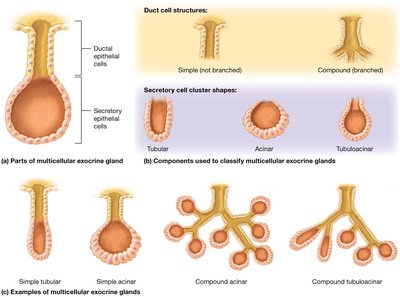
Structural Types of Exocrine Glands
Unicellular: Single cells, such as goblet cells, that secrete mucin.
Multicellular: Composed of a duct and a secretory unit; classified by duct structure (simple or compound) and shape (tubular, acinar, tubuloacinar).
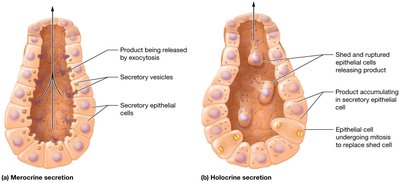
Modes of Secretion in Exocrine Glands
Merocrine: Secrete products by exocytosis (e.g., sweat, salivary glands).
Holocrine: Accumulate product until cell ruptures (e.g., sebaceous glands).
Apocrine: Accumulate product at the apex, which pinches off (e.g., mammary glands; debated in humans).
Connective Tissue
Overview and Functions
Connective tissue is the most abundant and widely distributed tissue type. It supports, protects, insulates, transports, stores energy, and binds organs. All connective tissues arise from embryonic mesenchyme and have varying degrees of vascularity and an extensive ECM.
Types of Connective Tissue
Connective Tissue Proper: Loose (areolar, adipose, reticular) and dense (regular, irregular, elastic).
Specialized Connective Tissue: Cartilage (hyaline, elastic, fibrocartilage), bone, and blood.
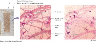
Loose (Areolar) Connective Tissue
Gel-like matrix with all three fiber types; contains fibroblasts, macrophages, mast cells, and some white blood cells. Functions in wrapping and cushioning organs, inflammation, and holding tissue fluid.
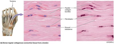
Dense Regular Connective Tissue
Parallel collagen fibers with few elastic fibers; fibroblasts are the main cell type. Provides strong attachment between structures (tendons, ligaments).
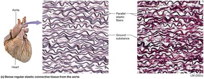
Dense Regular Elastic Connective Tissue
Contains parallel elastic fibers; allows tissues to recoil after stretching (found in large arteries).
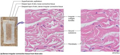
Dense Irregular Connective Tissue
Irregularly arranged collagen fibers; withstands tension from multiple directions (dermis, joint capsules).
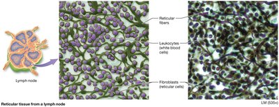
Reticular Connective Tissue
Network of reticular fibers in a loose ground substance; supports cells in lymphoid organs (lymph nodes, spleen, bone marrow).
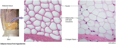
Adipose Tissue
Closely packed adipocytes with nucleus pushed to the side; stores energy, insulates, and cushions organs.
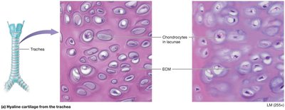
Cartilage
Hyaline Cartilage: Firm matrix with collagen fibers; supports, reinforces, and cushions. Found in embryonic skeleton, ends of long bones, trachea.
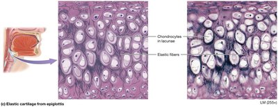
Elastic Cartilage: Similar to hyaline but with more elastic fibers; maintains shape and flexibility (external ear, epiglottis).
Fibrocartilage: Less firm matrix with thick collagen fibers; absorbs compressive shock (intervertebral discs, pubic symphysis, knee discs).
Bone (Osseous Tissue)
Hard, calcified matrix with collagen fibers; contains osteocytes in lacunae. Supports, protects, stores minerals, and forms blood cells.
Blood
Fluid matrix (plasma) with red and white blood cells and platelets; transports gases, nutrients, wastes, and other substances.
Muscle Tissue
Types of Muscle Tissue
Skeletal Muscle: Long, cylindrical, multinucleated cells with striations; voluntary movement.
Cardiac Muscle: Branching, striated, usually uninucleated cells with intercalated discs; involuntary, found in heart.
Smooth Muscle: Spindle-shaped, non-striated cells; involuntary, found in walls of hollow organs.
Nervous Tissue
Structure and Function
Nervous tissue consists of neurons and supporting cells (neuroglia). Neurons transmit electrical impulses, while neuroglia support and protect neurons. Main components include the cell body (soma), dendrites (input), and axon (output).
Additional info: For each tissue type, histological slides and diagrams are essential for visual identification and understanding of structure-function relationships. The images included above are directly relevant to the described tissues and their microscopic appearance.
