 Back
BackHistology: Structure and Function of Tissues
Study Guide - Smart Notes
 Tailored notes based on your materials, expanded with key definitions, examples, and context.
Histology: Structure and Function of Tissues
Tissue Overview
Histology is the study of tissues, which are groups of structurally and functionally related cells and their external environment. All tissues share two basic components: a discrete population of cells related in structure and function, and an extracellular matrix (ECM) that surrounds these cells.
Tissue: Group of cells and their external environment, working together for specific functions.
Extracellular Matrix (ECM): The material surrounding cells, providing structural and functional support.
Types of Tissues
The human body contains four primary tissue types, each with distinct roles:
Epithelial tissues: Tightly packed sheets of cells with little ECM; cover and line all body surfaces and cavities; specialized epithelia form glands.
Connective tissues: Connect all other tissues; largely composed of ECM; cells scattered throughout maintain the ECM.
Muscle tissues: Capable of generating force by contracting; little ECM between cells.
Nervous tissues: Store and transmit information; unique ECM.
Extracellular Matrix (ECM)
Functions and Components
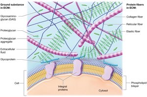
The ECM provides strength, holds cells in place, and regulates cell development, mitotic activity, and survival. It consists of ground substance and protein fibers.
Ground Substance: Extracellular fluid (ECF) composed of water and solutes, containing glycosaminoglycans (GAGs), proteoglycans, and cell-adhesion molecules (CAMs).
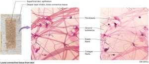
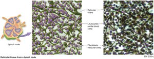
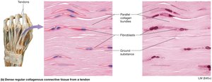
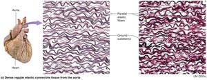
Protein Fibers: Collagen fibers (tensile strength), elastic fibers (stretch and recoil), and reticular fibers (support and scaffolding).
Epithelial Tissues
Functions
Epithelial tissues cover every internal and external body surface, acting as barriers and lining organs and cavities. Their functions include:
Protection: Shield underlying tissues from injury.
Immune Defense: Form physical barriers; contain immune cells.
Secretion: Form glands that produce substances like hormones and oils.
Transport: Selectively permeable membranes allow substances to cross into other tissues.
Sensation: Rich nerve supply; detect environmental changes.
Structure
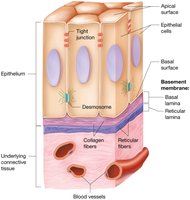
Epithelial tissues consist of tightly packed cells linked by tight junctions and desmosomes, making sheets of cells resistant to physical stresses. They are avascular and rely on diffusion for nutrients. The ECM is found beneath cells in a thin basement membrane, composed of basal lamina (collagen fibers and ground substance) and reticular lamina (reticular fibers and ground substance).

Classification of Epithelial Tissues
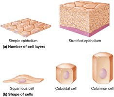
Epithelial tissues are classified by the number of cell layers and the shape of cells:
Simple epithelium: One cell layer.
Stratified epithelium: Multiple cell layers.
Cell shapes: Squamous (flat), cuboidal (cube-shaped), columnar (tall and rectangular).
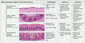
Simple Epithelia
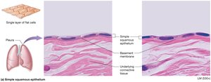
Simple squamous epithelium: Thin, single layer; rapid diffusion; found in air sacs of lungs, kidney tubules, and blood vessels.
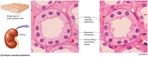
Simple cuboidal epithelium: Single layer of cube-shaped cells; rapid diffusion; found in renal tubules, respiratory passages, ducts of glands, and thyroid.
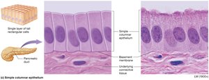
Simple columnar epithelium: Single layer of rectangular cells; often with microvilli or cilia; found in small intestine, uterine tubes, and respiratory tract.
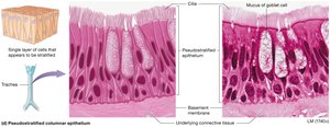
Pseudostratified columnar epithelium: Appears layered, but is one cell layer; nuclei at various heights; ciliated; found in respiratory tract and nasal cavity.
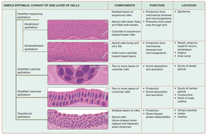
Stratified Epithelia
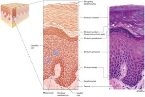
Keratinized stratified squamous epithelium: Apical layers are dead, filled with keratin; tough and resistant; found in skin.
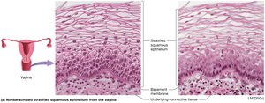
Nonkeratinized stratified squamous epithelium: Apical layers retain nuclei; moist surfaces; found in mouth, throat, esophagus, anus, and vagina.
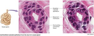
Stratified cuboidal epithelium: Two cell layers; lines ducts of sweat glands.
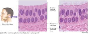
Stratified columnar epithelium: Few layers; apical layer is columnar; found in male urethra, cornea, and ducts of certain glands.
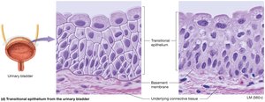
Transitional epithelium: Only in urinary system; apical cells flatten to allow stretching; found in kidney, ureters, bladder, and urethra.
Summary Tables of Epithelial Tissues
The following tables summarize the main types of epithelial tissues, their components, functions, and locations:
Type | Components | Function | Location |
|---|---|---|---|
Simple squamous | Single layer of flat cells | Rapid diffusion | Lungs, blood vessels |
Simple cuboidal | Single layer of cube-shaped cells | Absorption, secretion | Kidney tubules, glands |
Simple columnar | Single layer of tall cells | Absorption, secretion | Digestive tract, uterine tubes |
Pseudostratified columnar | Single layer, nuclei at different heights | Secretion, movement of mucus | Respiratory tract |
Type | Components | Function | Location |
|---|---|---|---|
Keratinized stratified squamous | Multiple layers, apical cells dead | Protection | Epidermis |
Nonkeratinized stratified squamous | Multiple layers, apical cells alive | Protection | Mouth, esophagus, vagina |
Stratified cuboidal | Two layers of cube-shaped cells | Protection, some absorption | Sweat gland ducts |
Stratified columnar | Two or more layers, apical columnar | Protection, some absorption | Salivary gland ducts, male urethra |
Transitional | Multiple layers, apical cells dome-shaped | Stretching | Urinary bladder |
Glandular Epithelium
Types of Glands
Glands are structures of epithelial origin that synthesize and secrete products. They are classified by shape and secretion mechanism:
Endocrine glands: Secrete hormones directly into bloodstream; systemic effects.
Exocrine glands: Release products onto surfaces or into ducts; local effects.
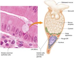
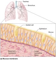
Goblet Cells
Goblet cells are the most common unicellular exocrine gland, found in digestive and respiratory tracts. They secrete mucus, a thick liquid that protects underlying epithelium.
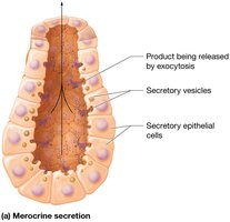
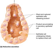
Modes of Secretion
Merocrine: Products released by exocytosis; most common (salivary and sweat glands).
Holocrine: Products accumulate and are released when cell ruptures and dies (sebaceous glands).
Apocrine: Portions of cytoplasm are pinched off with product; rare (mammary glands).
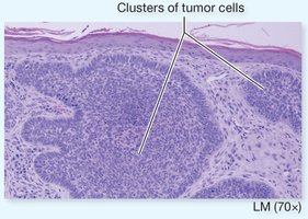
Pathology: Carcinoma
Carcinoma is cancer of epithelial tissue. The basement membrane acts as a barrier to prevent or slow the spread of carcinomas. Cancer cells produce enzymes that degrade the basement membrane, facilitating spread.
Connective Tissues
Structure and Functions
Connective tissue consists of cells and ECM. The ECM plays a major role in the function of each connective tissue type.
Connecting and binding: Anchor tissue layers and link organs.
Support: Bone and cartilage support body weight.
Protection: Bone protects organs; cartilage and fat absorb shock.
Transport: Blood transports substances throughout the body.
Connective Tissue Proper Cells
Fibroblasts: Most common; produce protein fibers and ground substance.
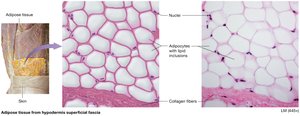
Adipocytes: Fat-storing cells.
Mast cells: Immune cells with granules of inflammatory mediators.
Phagocytes: Immune cells that ingest foreign substances; include macrophages and neutrophils.
Types of Connective Tissue Proper
Areolar (loose) connective tissue: Mostly ground substance; supports blood vessels and houses immune cells.
Adipose tissue: Fat-storing adipocytes; major energy reserve, insulation, shock absorption.
Reticular tissue: Mostly reticular fibers; supports small structures, forms nets in lymph nodes and spleen.
Dense regular connective tissue: Parallel collagen bundles; found in tendons and ligaments.
Dense regular elastic connective tissue: Parallel elastic fibers; found in walls of large blood vessels.
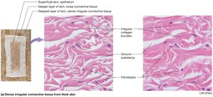
Dense irregular connective tissue: Disorganized collagen bundles; resists tension in all directions; found in dermis and around organs.
Specialized Connective Tissues
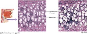
Cartilage
Elastic cartilage: Mostly elastic fibers; allows tissue to vibrate; found in external ear and larynx.
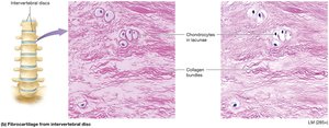
Fibrocartilage: Bundles of collagen fibers; great tensile strength; found in intervertebral discs.
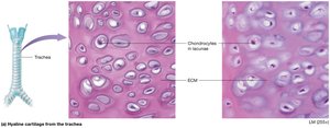
Hyaline cartilage: Most abundant; glossy appearance; found on ends of bones, respiratory tract, nose.
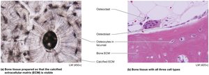
Bone
Bones have an ECM composed of organic (collagen fibers and ground substance) and inorganic (calcium phosphate crystals) components, making bone hard and strong.
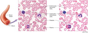
Blood
Blood is a fluid connective tissue with plasma as its ECM. It contains erythrocytes (red blood cells), leukocytes (white blood cells), and platelets (cell fragments).
Muscle Tissues
Structure and Physiology
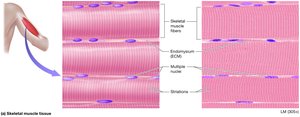
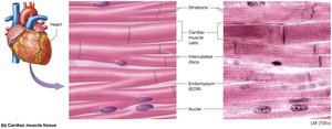
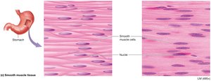
Muscle tissues are specialized for contraction, turning chemical energy (ATP) into mechanical energy. There are three types:
Skeletal muscle: Attached to skeleton; voluntary control; multinucleate cells.
Cardiac muscle: Only in heart; involuntary; striated; cells are short, branched, and usually uninucleate; intercalated discs allow coordinated contraction.
Smooth muscle: In walls of hollow organs; involuntary; cells are flattened with one nucleus; linked by gap junctions.
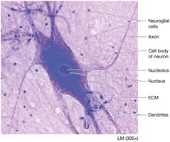
Nervous Tissues
Structure and Function
Nervous tissue makes up the brain, spinal cord, and nerves. It contains neurons and neuroglial cells, with a unique ECM.
Neuron: Excitable cell; cell body (soma), axon (transmits impulses), dendrites (receive impulses).
Neuroglial cells: Support neurons; anchor, monitor extracellular fluid, speed impulse transmission, circulate fluid.
Membranes
Types and Functions
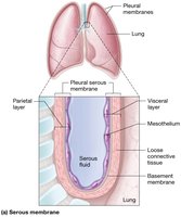
Membranes consist of a superficial epithelial layer resting on connective tissue. They anchor organs, serve as barriers, function in immunity, and secrete substances.
Serous membrane: Lines body cavities; two layers (parietal and visceral); produces serous fluid to reduce friction.
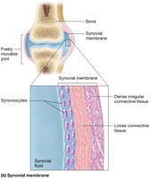
Synovial membrane: Lines joint cavities; two connective tissue layers; secretes synovial fluid for lubrication.
Mucous membrane: Lines passages opening to outside; contains goblet cells; produces mucus for protection.
Cutaneous membrane: Skin; outer layer of keratinized epithelium and underlying connective tissue.
Tissue Repair
Regeneration and Fibrosis
Tissue repair involves removing dead and damaged cells and replacing them. Some tissues regenerate (replace with same cell type), while others heal by fibrosis (scar tissue formation).
Regeneration: Skin, digestive tract, connective tissue proper, bone, and blood regenerate easily.
Fibrosis: Cartilage, cardiac muscle, skeletal muscle, and nervous tissue generally heal by fibrosis.
Factors affecting tissue repair include protein (collagen) production, vitamin C, and adequate blood supply.
