 Back
Human Physiology Exam Review – Osmosis, Neuron Structure, Action Potentials, Skeletal Muscle, and Endocrine Pathways
Study Guide - Smart Notes
 Tailored notes based on your materials, expanded with key definitions, examples, and context.
Q1. What is the % of water outside the cell?
Background
Topic: Osmosis and Cell Membrane Transport
This question tests your understanding of solute and solvent concentrations in solutions, specifically how to calculate the percentage of water when given the percentage of solute (glucose).
Key Terms:
Solute: The substance dissolved in a solution (here, glucose).
Solvent: The substance in which the solute is dissolved (here, water).
Percent composition: The percentage of each component in the solution.
Step-by-Step Guidance
Identify the percentage of glucose outside the cell (from the diagram: 80% glucose).
Recall that the total percentage of all components must add up to 100%.
Subtract the percentage of glucose from 100% to find the percentage of water outside the cell.
Try solving on your own before revealing the answer!
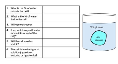
Final Answer: 20% water outside the cell
Since the solution outside the cell is 80% glucose, the remaining 20% must be water.
Q2. What is the % of water inside the cell?
Background
Topic: Osmosis and Cell Membrane Transport
This question is similar to Q1, but focuses on the internal environment of the cell.
Key Terms:
Intracellular: Inside the cell.
Solute and solvent percentages.
Step-by-Step Guidance
Identify the percentage of glucose inside the cell (from the diagram: 10% glucose).
Subtract the percentage of glucose from 100% to find the percentage of water inside the cell.
Try solving on your own before revealing the answer!
Final Answer: 90% water inside the cell
Since the cell contains 10% glucose, the remaining 90% is water.
Q3. Will osmosis occur?
Background
Topic: Osmosis
This question tests your understanding of osmosis, which is the movement of water across a semipermeable membrane from an area of higher water concentration to an area of lower water concentration.
Key Terms:
Osmosis: Movement of water across a membrane.
Semipermeable membrane: Allows water but not solute to pass.
Step-by-Step Guidance
Compare the water concentrations inside and outside the cell.
Determine if there is a difference in water concentration across the membrane.
If there is a difference, osmosis will occur.
Try solving on your own before revealing the answer!
Final Answer: Yes, osmosis will occur
There is a difference in water concentration, so water will move across the membrane.
Q4. If so, which way will water move (into or out of the cell)?
Background
Topic: Direction of Osmosis
This question tests your ability to predict the direction of water movement based on concentration gradients.
Key Terms:
Concentration gradient: Difference in concentration across a membrane.
Water moves from high to low concentration.
Step-by-Step Guidance
Identify which side of the membrane has higher water concentration (inside: 90%, outside: 20%).
Recall that water moves from high to low concentration.
Determine the direction of water movement based on these values.
Try solving on your own before revealing the answer!
Final Answer: Water will move out of the cell
Water moves from the area of higher concentration (inside) to lower concentration (outside).
Q5. Will the cell swell or shrink?
Background
Topic: Effects of Osmosis on Cell Volume
This question tests your understanding of how water movement affects cell size.
Key Terms:
Swelling: Cell gains water.
Shrinking: Cell loses water.
Step-by-Step Guidance
Recall the direction of water movement (out of the cell).
Predict the effect on cell volume when water leaves the cell.
Try solving on your own before revealing the answer!
Final Answer: The cell will shrink
As water leaves the cell, the cell loses volume and shrinks.
Q6. The cell is in what type of solution (hypertonic, isotonic, or hypotonic)?
Background
Topic: Tonicity
This question tests your understanding of solution types based on solute concentration relative to the cell.
Key Terms:
Hypertonic: Higher solute concentration outside the cell.
Isotonic: Equal solute concentration inside and outside.
Hypotonic: Lower solute concentration outside the cell.
Step-by-Step Guidance
Compare the solute concentration outside (80% glucose) to inside (10% glucose).
Determine which type of solution this represents.
Try solving on your own before revealing the answer!
Final Answer: Hypertonic solution
The solution outside the cell has a much higher solute concentration than inside, making it hypertonic.
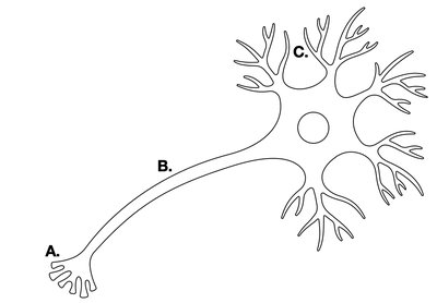
Q7. Use the image on the neuron to identify which segment (A, B, or C) the following channels/pumps will be located.
Background
Topic: Neuron Structure and Function
This question tests your knowledge of the anatomy of a neuron and the location of specific ion channels and pumps.
Key Terms:
Voltage-gated channels: Open in response to changes in membrane potential.
Chemical-gated channels: Open in response to binding of a chemical (e.g., neurotransmitter).
Segments of neuron: A (axon terminal), B (axon), C (dendrites/cell body).
Step-by-Step Guidance
Recall the function of each channel type and where they are typically found on a neuron.
Match each channel/pump to the correct segment (A, B, or C) based on its function.
Consider the direction of ion movement (ECF → ICF or ICF → ECF) for each channel.
Try solving on your own before revealing the answer!
Final Answer:
Voltage-gated potassium channels: B (axon), direction: ICF → ECF Voltage-gated calcium channels: A (axon terminal), direction: ECF → ICF Chemical-gated sodium channel: C (dendrites/cell body), direction: ECF → ICF
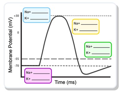
Q8. Below is a graph depicting change in membrane potential during an action potential. Within this graph, write the state of voltage-gated Na+ and K+ channels during each main phase of the action potential.
Background
Topic: Action Potential Phases
This question tests your understanding of the sequence of events during an action potential, including the opening and closing of sodium and potassium channels.
Key Terms:
Depolarization: Na+ channels open, Na+ enters cell.
Repolarization: K+ channels open, K+ leaves cell.
Resting state: Both channels closed.
Hyperpolarization: K+ channels remain open briefly.
Step-by-Step Guidance
Identify the phases of the action potential on the graph (resting, depolarization, repolarization, hyperpolarization).
For each phase, determine the state (open/closed) of Na+ and K+ channels.
Write the state for each channel in the corresponding box on the graph.
Try solving on your own before revealing the answer!
Final Answer:
Resting: Na+ closed, K+ closed Depolarization: Na+ open, K+ closed Repolarization: Na+ closed, K+ open Hyperpolarization: Na+ closed, K+ open (briefly)
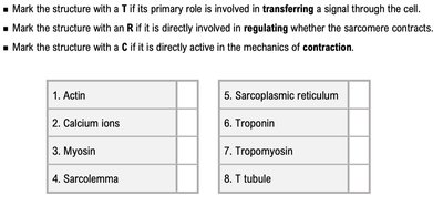
Q9. Mark the structure with a T if its primary role is involved in transferring a signal through the cell. Mark the structure with an R if it is directly involved in regulating whether the sarcomere contracts. Mark the structure with a C if it is directly active in the mechanics of contraction.
Background
Topic: Skeletal Muscle Structure and Function
This question tests your understanding of the roles of various muscle cell structures in contraction, regulation, and signal transmission.
Key Terms:
Transferring (T): Structures that transmit signals (e.g., T tubule, sarcolemma).
Regulating (R): Structures that control contraction (e.g., calcium ions, troponin).
Contracting (C): Structures directly involved in contraction (e.g., actin, myosin).
Step-by-Step Guidance
Review the function of each listed structure.
Assign T, R, or C based on the primary role of each structure.
Mark each structure accordingly.
Try solving on your own before revealing the answer!
Final Answer:
Actin: C Calcium ions: R Myosin: C Sarcolemma: T Sarcoplasmic reticulum: T Troponin: R Tropomyosin: R T tubule: T
Q10. The diagram below represents a typical hypothalamus-pituitary endocrine control pathway. Identify each labeled component of the pathway.
Background
Topic: Endocrine Physiology
This question tests your understanding of the organization and function of the hypothalamus-pituitary axis and negative feedback regulation.
Key Terms:
Hypothalamus: Integrating center.
Anterior pituitary: Sensor and effector.
Target gland: Effector.
Negative feedback: Regulation mechanism.
Step-by-Step Guidance
Identify the role of each labeled box (A–F) in the pathway.
Recall the sequence: hypothalamus → pituitary → target gland → hormone effect.
Match each label to its corresponding structure or function.
Try solving on your own before revealing the answer!
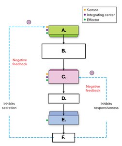
Final Answer:
A: Hypothalamus B: Releasing hormone C: Anterior pituitary D: Stimulating hormone E: Target gland F: Hormone released by target gland
