 Back
Immunity: Host Defenses and the Immune Response
Study Guide - Smart Notes
 Tailored notes based on your materials, expanded with key definitions, examples, and context.
Immunity and Host Defenses
Overview of Major Host Defenses
The human body is protected from pathogens by a complex system of host defenses, which are divided into innate (nonspecific) and acquired (specific) immunity. These defenses operate at multiple levels to prevent infection and eliminate invading microbes.
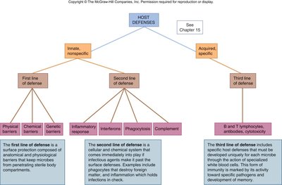
Innate (Nonspecific) Immunity: Provides immediate, general protection against pathogens. Includes physical, chemical, and genetic barriers, as well as cellular responses like inflammation and phagocytosis.
Acquired (Specific) Immunity: Develops after exposure to specific antigens and involves lymphocytes (B and T cells) and the production of antibodies. This system has memory, allowing for a faster and stronger response upon re-exposure to the same pathogen.
First Line of Defense: Physical, chemical, and genetic barriers that prevent pathogen entry.
Second Line of Defense: Inflammatory response, interferons, phagocytosis, and complement system that act when pathogens breach the first line.
Third Line of Defense: Specific immune responses involving B and T lymphocytes, antibodies, and cytotoxicity.
Inflammatory Response
Classic Signs and Symptoms
The inflammatory response is a key component of innate immunity, characterized by five classic signs: redness, warmth, swelling, pain, and loss of function. These signs result from increased blood flow, vascular permeability, and the accumulation of immune cells at the site of injury or infection.
Redness (Rubor): Caused by increased circulation and vasodilation in injured tissues due to chemical mediators.
Warmth (Calor): Heat produced by increased blood flow to the area.
Swelling (Tumor): Edema from fluid, white blood cells, microbes, and debris accumulating in tissues.
Pain (Dolor): Stimulation of nerve endings by chemicals and swelling.
Loss of Function: Impaired movement or use of the affected area due to swelling and pain.
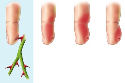
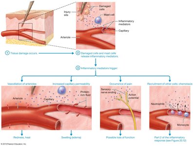
Example: A cut or puncture wound triggers inflammation, resulting in redness, swelling, and pain at the site.
Phagocytosis
Mechanism and Steps
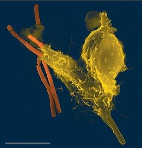
Phagocytosis is the process by which specialized cells (phagocytes) engulf and destroy pathogens and debris. This is a crucial part of the innate immune response.
Chemotaxis: Phagocytes are attracted to the site of infection by chemical signals.
Adhesion: Phagocytes adhere to pathogens using receptors that recognize pathogen-associated molecular patterns (PAMPs).
Engulfment: The pathogen is engulfed into a phagocytic vacuole (phagosome).
Phagolysosome Formation: The phagosome fuses with a lysosome, forming a phagolysosome where enzymes and reactive oxygen species destroy the pathogen.
Destruction and Release: Pathogen is digested, and residual debris is expelled from the cell.
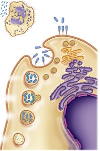
Key Enzymes: Lysozyme, DNase, RNase, proteases, and peroxidase are involved in the breakdown of pathogens.
Reactive Oxygen Products: Superoxide (), hydrogen peroxide (), singlet oxygen (), hydroxyl ion (), and hypochlorite ion () contribute to microbial killing.
Immunoglobulins (Antibodies)
Classes and Functions
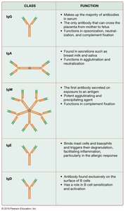
Immunoglobulins (Ig) are glycoproteins produced by B cells that recognize and bind to specific antigens. There are five main classes, each with distinct functions in the immune response.
Class | Function |
|---|---|
IgG | Majority of antibodies in serum; only antibody to cross the placenta; functions in opsonization, neutralization, and complement fixation. |
IgA | Found in secretions (e.g., breast milk, saliva); protects mucosal surfaces. |
IgM | First antibody secreted upon antigen exposure; potent agglutinating and complement-fixing agent. |
IgE | Binds mast cells and basophils; involved in allergic responses and defense against parasites. |
IgD | Found on B cell surfaces; involved in B cell sensitization and activation. |
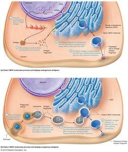
Antibody Structure: Antibodies have variable regions (antigen-binding sites), constant regions, light and heavy chains, and complement-binding sites.
Primary and Secondary Immune Responses
Comparison and Characteristics
The immune system responds differently upon first and subsequent exposures to an antigen. The primary response is slower and produces less antibody, while the secondary response is faster and stronger due to immunological memory.
Characteristic | Primary Immune Response | Secondary Immune Response |
|---|---|---|
Lag phase | 4–5 days | 1–3 days |
Time until antibody peak | 7–14 days | 3–5 days |
Primary antibody | IgM | IgG |
Duration of response | 14–21 days | 28 days and beyond |
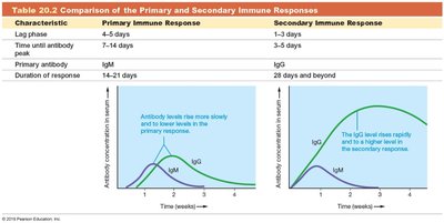
Example: Vaccination primes the immune system, so upon real infection, the secondary response is rapid and effective.
Summary Table: Humoral vs. Cellular Immunity
Adaptive immunity is divided into humoral (antibody-mediated) and cellular (cell-mediated) responses. Humoral immunity involves B cells and antibody production, while cellular immunity involves T cells targeting infected or abnormal cells.
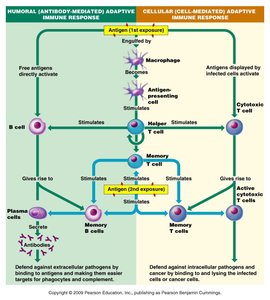
Humoral Immunity: Defends against extracellular pathogens via antibodies produced by plasma cells (B cells).
Cellular Immunity: Defends against intracellular pathogens and abnormal cells via cytotoxic T cells.
Memory Cells: Both B and T cells generate memory cells for faster response upon re-exposure to the same antigen.
