 Back
BackIntegumentary System: Structure, Functions, and Clinical Aspects
Study Guide - Smart Notes
 Tailored notes based on your materials, expanded with key definitions, examples, and context.
Integumentary System Overview
Components of the Integumentary System
The integumentary system is a complex organ system that includes the skin and its appendages. It serves as the body's primary interface with the external environment and provides multiple protective and regulatory functions.
Skin (cutaneous membrane): The largest organ of the body, consisting of multiple layers.
Skin appendages: Includes sweat glands, oil glands, hair, and nails.
Functions of the Integumentary System
The integumentary system performs several essential functions to maintain homeostasis and protect the body.
Insulation and cushioning: Protects deeper organs from physical trauma.
Protection: Guards against mechanical, chemical, thermal, UV, and microbial damage, as well as desiccation.
Synthesizes vitamin D: Facilitates vitamin D production when exposed to sunlight.
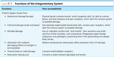
Table: Functions of the Integumentary System
Functions | How accomplished |
|---|---|
Protects deeper tissues from mechanical, chemical, microbe, UV, thermal, and desiccation damage | Physical barrier (keratin, fat, pressure/pain receptors), keratinized cells, acid mantle, phagocytes, melanin, heat/cold/pain receptors, water-resistant glycolipids |
Aids in body heat loss or retention | Heat loss: sweat glands activation and blood flushing; Heat retention: limiting blood flow to skin capillaries |
Aids in excretion of urea and uric acid | Contained in sweat gland perspiration |
Synthesizes vitamin D | Modified cholesterol molecules in skin converted to vitamin D in sunlight |
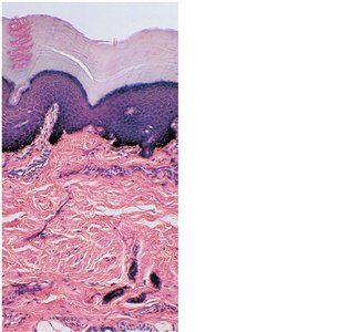
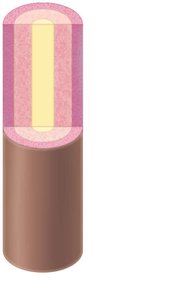
Structure of the Skin
Layers of the Skin
The skin is composed of two primary layers: the epidermis and the dermis, with a subcutaneous layer beneath.
Epidermis: The outer layer, made of stratified squamous epithelium. It is avascular and consists of five strata. Keratinocytes produce keratin, providing toughness and water resistance.
Dermis: The deeper layer, composed of connective tissue. It contains blood vessels, nerves, and skin appendages.
Hypodermis (subcutaneous tissue): Not technically part of the skin, but anchors skin to underlying tissues and stores fat.
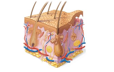
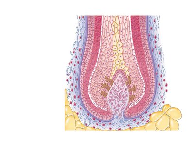
Microscopic Features of the Epidermis
The epidermis contains several specialized cells and distinct layers:
Keratinocytes: Most common cell, produces keratin.
Melanocytes: Produce melanin pigment.
Merkel cells: Sensory receptors for touch.
Epidermal dendritic cells: Immune defense.
Stratum basale: Deepest layer, site of cell division.
Stratum spinosum: Contains pre-keratin filaments.
Stratum granulosum: Cells flatten, organelles deteriorate.
Stratum corneum: Outermost layer, dead keratinized cells.
Microscopic Features of the Dermis
The dermis is divided into two layers:
Papillary layer: Contains dermal papillae, capillaries, and sensory receptors.
Reticular layer: Contains dense connective tissue, blood vessels, sweat and oil glands.
Skin Color
Pigments Contributing to Skin Color
Skin color is determined by three main pigments:
Melanin: Yellow, reddish brown, or black pigment produced by melanocytes.
Carotene: Orange-yellow pigment from vegetables.
Hemoglobin: Red pigment from blood cells; oxygen content affects redness.
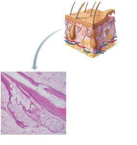
Appendages of the Skin
Cutaneous Glands
Cutaneous glands are exocrine glands that include sebaceous and sweat glands.
Sebaceous (oil) glands: Produce sebum, which softens skin, prevents hair brittleness, and kills bacteria. Most ducts empty into hair follicles; activated at puberty.
Sweat glands: Include eccrine and apocrine glands. Eccrine glands open to skin surface and regulate temperature; apocrine glands empty into hair follicles in armpit/genital regions and function at puberty.
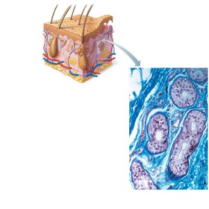
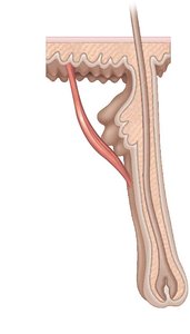
Hair
Hair is produced by hair follicles and consists of hard keratinized epithelial cells. Melanocytes provide pigment for hair color. Hair grows in the matrix of the hair bulb in the stratum basale.
Root: Enclosed in the follicle.
Shaft: Projects from the skin surface.
Arrector pili muscle: Causes hair to stand up.
Nails
Nails are heavily keratinized, scalelike modifications of the epidermis. The stratum basale extends beneath the nail bed and is responsible for nail growth. Nails lack pigment, making them colorless.
Homeostatic Imbalances of Skin
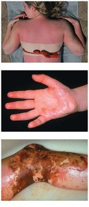
Burns
Burns are classified by their depth and severity. The extent of a burn is estimated using the rule of nines, which divides the body into regions representing 9% (or multiples) of total body surface area.
First-degree burn: Only epidermis is damaged; skin is red and swollen.
Second-degree burn: Epidermis and superficial dermis damaged; skin is red, painful, and blistered.
Third-degree burn: Destroys epidermis and dermis; area is painless, requires skin grafts.
Fourth-degree burn: Extends into deeper tissues; may require surgery or amputation.
Skin Cancer
Skin cancer is a common homeostatic imbalance. The three main types are:
Basal cell carcinoma: Least malignant, most common; arises from stratum basale cells.
Squamous cell carcinoma: Induced by UV exposure; arises from stratum spinosum cells.
Malignant melanoma: Most deadly; arises from melanocytes, rapidly metastasizes.
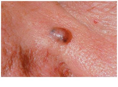
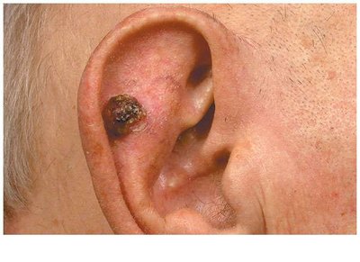
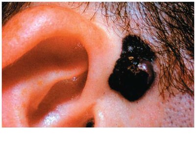
ABCDE Rule for Melanoma Detection
A = Asymmetry: Two sides of mole do not match.
B = Border irregularity: Borders are not smooth.
C = Color: Multiple colors present.
D = Diameter: Larger than 6 mm.
E = Evolution: Changes in any of the above characteristics.
Developmental Aspects of Skin and Body Membranes
Fetal and Neonatal Skin
During fetal development, lanugo (downy hair) covers the body by the fifth or sixth month but disappears by birth. Vernix caseosa, an oily covering, is apparent at birth and protects the skin.
Additional info: Academic context was added to clarify the structure and function of skin layers, appendages, and clinical aspects. All images included are directly relevant to the adjacent explanations.
