 Back
BackIntroduction to Human Physiology: The Human Body, Cells, and Homeostasis
Study Guide - Smart Notes
 Tailored notes based on your materials, expanded with key definitions, examples, and context.
The Human Body: An Orientation
Definition and Scope of Anatomy and Physiology
Anatomy is the study of the structure of body parts, including their names, locations, and roles. Physiology focuses on the function of these parts, explaining how and why they work as they do. Together, these disciplines provide a comprehensive understanding of the human body.
Anatomy: Structure, location, and identification of organs.
Physiology: Function and mechanisms of body parts.
Homeostasis: The body's ability to maintain a stable internal environment despite external changes.
The internal environment refers to the extracellular fluid (ECF), which surrounds cells and acts as a buffer between cells and the external world.
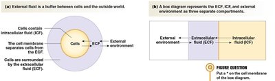
Cells contain intracellular fluid (ICF), and the cell membrane separates the ICF from the ECF. The ECF itself is composed of the liquid portion of blood (plasma) and interstitial fluid.
Homeostatic Balances
The body regulates several key variables to maintain homeostasis:
Glucose levels
Oxygen levels
Temperature
Acidity (pH)
Waste levels (e.g., carbon dioxide, ammonia)
Electrolytes and water levels
Pressure and volume of fluids
Multiple organ systems work together to regulate these factors and maintain the preferred environment for cellular function.

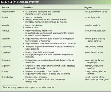
Major Organ Systems
The human body is organized into several organ systems, each with specific functions essential for survival and homeostasis.
System | Functions | Organs |
|---|---|---|
Integumentary | Barrier to pathogens, prevents water loss | Skin, subcutaneous tissue |
Skeletal | Supports body, protects organs, forms blood | Bones, ligaments |
Muscular | Moves skeleton, produces heat | Muscles, tendons |
Nervous | Regulates body functions, interprets sensation | Brain, nerves, eyes, ears |
Endocrine | Regulates metabolism, growth, reproduction | Thyroid, pituitary, adrenals |
Circulatory | Transports oxygen, nutrients, wastes | Heart, blood, arteries, veins |
Lymphatic | Returns tissue fluid to blood, immunity | Spleen, lymph nodes |
Respiratory | Exchanges oxygen and carbon dioxide | Lungs, trachea, larynx |
Digestive | Breaks down food, absorbs nutrients | Stomach, colon, liver, pancreas |
Urinary | Removes waste, regulates blood volume | Kidneys, urinary bladder |
Reproductive | Produces gametes, supports embryo | Ovaries, uterus, testes, prostate |
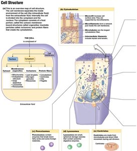
Cells: The Living Units
Basic Cell Structure
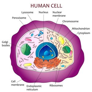
Cells are the fundamental units of life. Each cell is surrounded by a plasma membrane, contains a nucleus, and has cytoplasm filled with organelles and cytosol. The cytoskeleton provides structural support and facilitates movement.
Cell (plasma) membrane: Encloses the cell, regulates entry and exit of substances.
Nucleus: Contains genetic material (DNA) and the nucleolus (site of ribosome synthesis).
Cytoplasm: Includes organelles (e.g., endoplasmic reticulum, Golgi apparatus, lysosomes, mitochondria), cytosol, and cytoskeleton.
Ribosomes: Sites of protein synthesis (not membrane-bound).
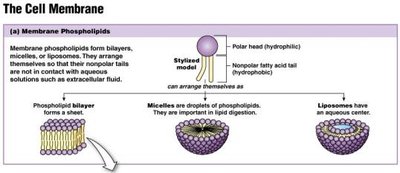
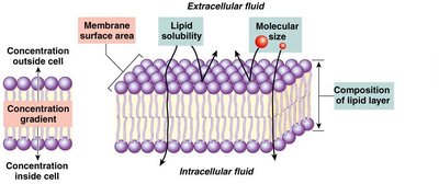
The Cell Membrane
The cell membrane is a phospholipid bilayer that acts as a selective barrier, controlling the movement of substances into and out of the cell. Hydrophobic molecules can pass through the lipid interior, while hydrophilic molecules require specific transport mechanisms.
Hydrophobic molecules: Can dissolve through the lipid bilayer (e.g., O2, CO2, steroid hormones).
Hydrophilic molecules: Require channels or transporters (e.g., ions, glucose, amino acids).
Major Cell Organelles
Nucleus: Double-membraned, contains DNA and nucleolus.
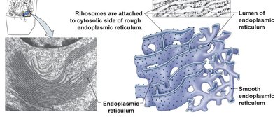
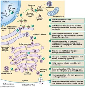
Endoplasmic Reticulum (ER): Rough ER (with ribosomes) synthesizes proteins; Smooth ER synthesizes lipids and detoxifies chemicals.
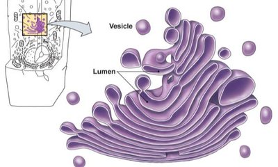
Golgi Apparatus: Modifies, sorts, and packages proteins and lipids for delivery.
Mitochondria: Site of ATP (energy) production.
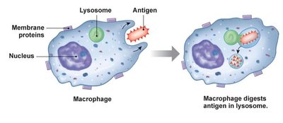
Lysosomes: Contain digestive enzymes for breaking down waste and foreign material.
Cytoskeleton: Provides structure and facilitates movement (microtubules, microfilaments, intermediate filaments).
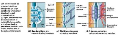
Cell Junctions
Cells are connected by specialized junctions that facilitate communication and maintain tissue integrity:
Desmosomes: Anchor cells together.
Gap junctions: Allow direct communication between cells.
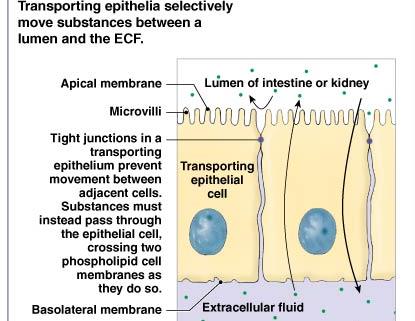
Tight junctions: Prevent movement of substances between cells.
Membranes and Cell Communication
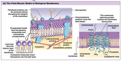
Membrane Structure and Function
The cell membrane is composed of a phospholipid bilayer with embedded proteins, cholesterol, glycoproteins, and glycolipids. It acts as a barrier to most polar molecules and all ions, but is permeable to hydrophobic and small polar molecules. Membrane proteins serve structural, enzymatic, receptor, and transporter functions.
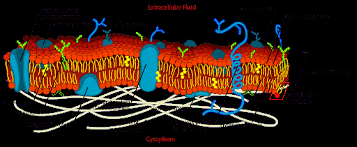
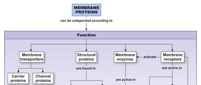
Membrane Proteins
Membrane proteins are categorized by function:
Structural proteins: Maintain cell shape and connect cells.
Enzymes: Catalyze reactions at the membrane surface.
Receptors: Receive and transmit signals.
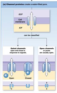
Transporters: Move substances across the membrane (carriers and channels).
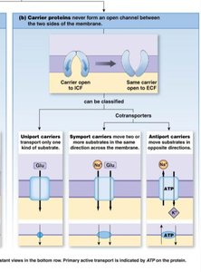
Transport Across Membranes
Transport can be passive (no energy required) or active (requires energy):
Simple diffusion: Movement down a concentration gradient without assistance.
Facilitated diffusion: Movement down a gradient via a protein transporter.
Active transport: Movement against a gradient, requiring energy (often ATP).
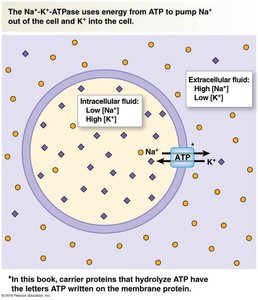
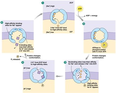
Active Transport: The Na+/K+ Pump
The sodium-potassium pump (Na+/K+-ATPase) is a primary active transporter that moves Na+ out of and K+ into the cell, maintaining essential ion gradients for cell function.
Uses ATP to transport 3 Na+ out and 2 K+ in per cycle.
Critical for nerve impulse transmission and muscle contraction.
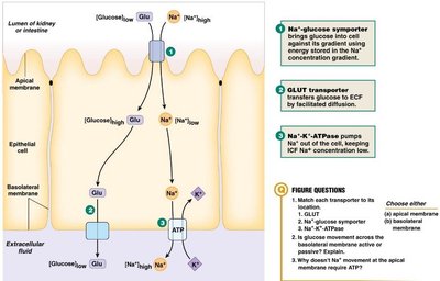
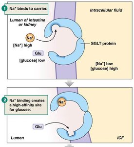
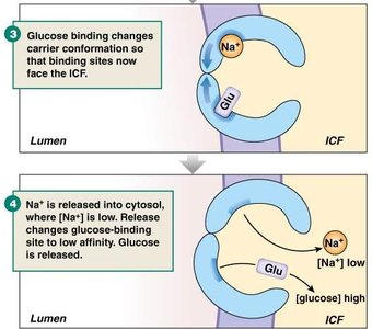
Glucose Transport
Glucose is transported into cells via symporters that use the Na+ gradient (secondary active transport), and then exits into the bloodstream via facilitated diffusion.
Na+-glucose symporter: Moves glucose into the cell using Na+ gradient energy.
GLUT transporter: Facilitates glucose exit into the bloodstream.
Additional info:
Cells communicate via short- and long-distance signaling, including gap junctions, paracrine, autocrine, endocrine, and synaptic mechanisms.
Homeostasis is maintained primarily through negative feedback mechanisms, with positive feedback occurring in specific cases (e.g., childbirth, blood clotting).
