 Back
BackIntroduction to the Nervous System and Nervous Tissue
Study Guide - Smart Notes
 Tailored notes based on your materials, expanded with key definitions, examples, and context.
Introduction to the Nervous System
Overview
The nervous system is a complex network responsible for coordinating all body activities. It is divided into the central nervous system (CNS) and the peripheral nervous system (PNS), each with distinct structures and functions.
Anatomy Review
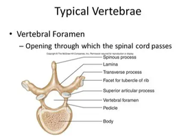
Vertebral Foramen
The vertebral foramen is the opening in each vertebra through which the spinal cord passes, providing protection and structural support.
The Great Divide: CNS and PNS
Central Nervous System (CNS)
Brain: Contains approximately 100 billion neurons, located in the skull, and controls all body functions.
Spinal Cord: Contains about 100 million neurons, passes through the foramen magnum and vertebral foramen, and serves as a communication pathway between the brain and the rest of the body.
Peripheral Nervous System (PNS)
Cranial Nerves: 12 pairs, connect directly to the brain.
Spinal Nerves: 31 pairs, connect to the spinal cord and branch throughout the body.
Functional Divisions of the Nervous System
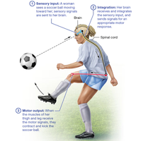
Sensory, Integrative, and Motor Functions
The nervous system performs three main functions:
Sensory Input: Gathering information about internal and external environments.
Integration: Analyzing and interpreting sensory information.
Motor Output: Initiating actions in response to integration.
Nervous Tissue Basics
Cellular Composition
Neurons: The primary signaling cells, responsible for transmitting electrical impulses.
Neuroglia: Supportive cells that maintain the environment, protect neurons, and assist in their function.
Extracellular Matrix: Provides structural support.
Axons and Dendrites: Specialized extensions for communication.
The Neuron
Structure and Function
Cell Body (Soma): Metabolically active, contains the nucleus and organelles.
Dendrites: Receive input from other neurons; their branching increases receptive surface area.
Axon: Single extension that transmits signals away from the cell body; may branch into axon collaterals and telodendria.
Axonal Transport
Slow Axonal Transport: Moves cytoskeletal proteins at 1–3 mm/day.
Fast Axonal Transport: Uses motor proteins and ATP to move materials at up to 200 mm/day.
Direction: Anterograde (toward axon terminal), Retrograde (toward cell body).
Functional Regions of a Neuron
Receptive Region: Dendrites and cell body; receive signals.
Conducting Region: The axon; transmits signals.
Secretory Region: Axon terminals; release neurotransmitters.
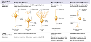
Structural Classes of Neurons
Classification and Features
Neurons are classified based on the number and arrangement of their processes:
Structural Class | Features | Typical Function | Location |
|---|---|---|---|
Multipolar Neurons | One axon, two or more dendrites | Motor (efferent) neurons, interneurons | Most neurons in CNS, motor neurons in PNS |
Bipolar Neurons | One axon, one dendrite | Sensory (afferent) neurons | Special sense organs (retina, olfactory epithelium) |
Pseudounipolar Neurons | Single process splits into two axons | Sensory (afferent) neurons | Sensory neurons in PNS (touch, pain, vibration) |
Neuroglia
Supportive Cells
Hold neurons together, maintain the extracellular environment, protect neurons, and fill gaps after injury.
Myelin Sheath and Matter Types
Myelination
Myelin Sheath: Lipid-rich covering that insulates axons, preventing current leakage and speeding up signal transmission.
White Matter: Regions with myelinated axons.
Gray Matter: Regions with unmyelinated axons and cell bodies.
Regeneration in the Nervous System
Steps of Axonal Regeneration
Distal axon and myelin sheath degenerate.
Growth processes form from the proximal axon.
Neurolemmocytes and basal lamina form a regeneration tube.
A single growth process enters the tube.
The axon reconnects with the target cell.
Membrane Potentials and Channels
Resting Membrane Potential
Difference in charge across the neuron's membrane at rest, typically around -70 mV.
Maintained by sodium-potassium pumps and leak channels.
Electrochemical Gradient
Combined effect of electrical and chemical gradients that drive ion movement across membranes.
Local and Action Potentials
Local (Graded) Potentials
Small changes in membrane potential due to stimulation.
Depolarization: Positive charges enter, making the membrane less negative.
Hyperpolarization: Positive charges exit or negative charges enter, making the membrane more negative.
Action Potentials
Large, rapid changes in membrane potential that propagate along the axon.
Involve voltage-gated sodium and potassium channels.
Voltage-Gated Channel States
Resting State: Inactivation gate open, activation gate closed.
Activated State: Both gates open.
Inactivated State: Inactivation gate closed, activation gate open.
Neuronal Synapses
Types of Synapses
Electrical Synapses: Direct current flow via gap junctions; bidirectional and nearly instantaneous.
Chemical Synapses: Use neurotransmitters to transmit signals across a synaptic cleft; more common and efficient.
Events at a Chemical Synapse
Action potential triggers calcium channels to open in the presynaptic terminal.
Calcium influx causes synaptic vesicles to release neurotransmitters into the synaptic cleft.
Neurotransmitters bind to receptors on the postsynaptic neuron, opening ion channels and generating a local potential.
Termination of Synaptic Transmission
Diffusion and absorption
Degradation in the synaptic cleft
Reuptake into the presynaptic neuron
Neurotransmitters
Major Types and Functions
Acetylcholine: Involved in muscle activation and autonomic functions.
Biogenic Amines (Monoamines): Include norepinephrine, epinephrine, dopamine, serotonin, and histamine; regulate mood, cognition, and autonomic functions.
Amino Acid Neurotransmitters: Glutamate (excitatory), GABA (inhibitory in brain), Glycine (inhibitory in spinal cord).
Neuropeptides: Substance P (pain), opioids (pain relief), neuropeptide Y (hunger).
Neuromodulation
Neurotransmitters that modulate neuron activity over longer periods, often through metabolic receptors and signaling cascades.
Clinical Applications and Disorders
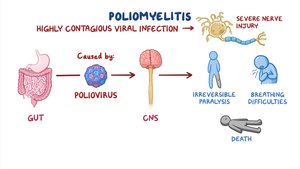
Poliomyelitis
Poliovirus enters the CNS via retrograde axonal transport, causing severe nerve injury, paralysis, and potentially death. Other pathogens (herpes simplex, rabies, tetanus) use similar mechanisms.
Brain Fog
Microglia can reduce oligodendrocyte precursors and myelin, slowing impulse conduction.
Psychiatric Disorders
Schizophrenia: Excess dopamine; treated with dopamine receptor blockers.
Depressive Disorders: Deficiency in serotonin, norepinephrine, or dopamine; treated with SSRIs and related drugs.
Anxiety Disorders: Abnormal norepinephrine, serotonin, and GABA; treated with drugs enhancing GABA or modulating norepinephrine.
Bipolar Disorder: Treated with drugs that block sodium channels to reduce action potential generation.
Sample Review Questions
What is resting membrane potential?
Is the concentration of sodium ions greater in the cytosol or the extracellular fluid?
What is an electrochemical gradient?
What is a negative feedback loop? Example?
What is a positive feedback loop? Example?
What takes place during an action potential?
Which cells form cerebrospinal fluid? (Answer: Ependymal cells)
The influx of positive ions that makes the membrane potential less negative is called: Depolarization
Which of the following is an inhibitory neurotransmitter? (Answer: Glycine)
