 Back
BackIntroduction to the Nervous System and Nervous Tissue
Study Guide - Smart Notes
 Tailored notes based on your materials, expanded with key definitions, examples, and context.
Nervous System & Nervous Tissue
Anatomy Review
The nervous system is a complex network responsible for coordinating the body's activities. It is divided into the central nervous system (CNS) and the peripheral nervous system (PNS). The CNS consists of the brain and spinal cord, while the PNS includes cranial and spinal nerves that connect the CNS to the rest of the body.
The Great Divide: CNS vs. PNS
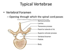
Central Nervous System (CNS): Includes the brain (100 billion neurons, located in the skull, controls all body tasks) and spinal cord (100 million neurons, passes through the foramen magnum and vertebral foramen, connects brain to body).
Peripheral Nervous System (PNS): Composed of cranial nerves (12 pairs, connect to the brain) and spinal nerves (31 pairs, connect to the spinal cord).
Functional Divisions of the Nervous System
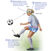
The nervous system is functionally divided into sensory, integrative, and motor divisions:
Sensory Functions: Gather information about internal and external environments.
Integrative Functions: Analyze and interpret sensory stimuli.
Motor Functions: Actions performed in response to integration.
Nervous Tissue Basics
Cellular Composition
Neurons: Excitable cells that transmit electrical signals.
Neuroglia: Supportive cells that maintain the environment, protect neurons, and assist in their function.
Extracellular Matrix: Provides structural support.
Axons and Dendrites: Specialized processes for signal transmission and reception.
The Neuron
Cell Body (Soma): Metabolically active, contains cytoplasm and organelles for protein synthesis.
Dendrites: Receive input from other neurons; their branching increases receptive surface area and is pruned during maturation.
Axon: Single process that transmits signals away from the cell body; may branch into axon collaterals and telodendria ending in axon terminals or synaptic knobs.
Axonal Transport
Slow Axonal Transport: Moves cytoskeletal proteins at 1-3 mm/day.
Fast Axonal Transport: Uses motor proteins and ATP to move materials at 200 mm/day.
Direction: Anterograde (toward axon terminal), Retrograde (away from axon terminal).
Functional Regions of a Neuron
Receptive Region: Dendrites and cell body; receive signals.
Conducting Region: The axon; transmits signals.
Secretory Region: Axon terminals; release neurotransmitters.
Structural Classes of Neurons
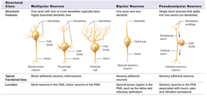
Neurons are classified based on the number and arrangement of their processes:
Multipolar Neurons: One axon, multiple dendrites; most common in CNS.
Bipolar Neurons: One axon, one dendrite; found in special sensory organs.
Pseudounipolar Neurons: Single process splits into two branches; general sensory neurons.
Neuroglia
Support and protect neurons.
Maintain the extracellular environment.
Fill gaps when neurons die.
Myelin Sheath
Insulates axons, preventing current leakage and speeding up signal transmission.
High lipid content makes it an excellent insulator, similar to rubber around a wire.
White Matter: Myelinated axons.
Gray Matter: Unmyelinated axons and cell bodies.
Regeneration in the PNS
Distal axon and myelin sheath degenerate.
Growth processes form from the proximal axon.
Neurolemmocytes and basal lamina form a regeneration tube.
A single growth process enters the tube.
Axon reconnects with the target cell.
Neuronal Physiology
Resting Membrane Potential
Difference in charge across the neuron's membrane at rest, typically -70 mV.
Maintained by sodium-potassium pumps and leak channels.
Sodium ions are more concentrated in the extracellular fluid; potassium ions are higher in the cytosol.
Electrochemical Gradient: Combination of electrical and chemical forces driving ion movement.
Local Potentials
Small, graded changes in membrane potential due to stimulation.
Depolarization: Positive charges enter, making the membrane less negative.
Hyperpolarization: Positive charges exit or negative charges enter, making the membrane more negative.
Action Potentials
Large, rapid changes in membrane potential that propagate along the axon.
Involve voltage-gated sodium and potassium channels cycling through resting, activated, and inactivated states.
Voltage-Gated Channels
Resting State: Inactivation gate open, activation gate closed.
Activated State: Both gates open.
Inactivated State: Inactivation gate closed, activation gate open.
Neuronal Synapses
Overview
Presynaptic Neuron: Sends the message.
Postsynaptic Neuron: Receives the message.
Synaptic Transmission: Transfer of signals between neurons via chemical or electrical means.
Electrical Synapses
Cells are connected by gap junctions, allowing direct current flow.
Transmission is bidirectional and nearly instantaneous.
Chemical Synapses
More common and efficient than electrical synapses.
Key structures: synaptic vesicles (store neurotransmitters), synaptic cleft, neurotransmitter receptors.
Events at a Chemical Synapse
Action potential triggers calcium channels to open in the presynaptic terminal.
Calcium influx causes vesicles to release neurotransmitters into the synaptic cleft.
Neurotransmitters bind to postsynaptic receptors, opening ion channels and generating a local potential.
Termination of Synaptic Transmission
Diffusion and absorption of neurotransmitter.
Degradation by enzymes in the synaptic cleft.
Reuptake into the presynaptic neuron.
Neurotransmitters and Neuromodulation
Major Neurotransmitter Classes
Acetylcholine: Involved in muscle activation and autonomic functions.
Biogenic Amines (Monoamines): Include norepinephrine, epinephrine, dopamine, serotonin, and histamine. Regulate mood, cognition, and autonomic functions.
Amino Acid Neurotransmitters: Glutamate (main excitatory NT in the brain), GABA (main inhibitory NT in the brain), Glycine (main inhibitory NT in the spinal cord).
Neuropeptides: Substance P (pain and temperature), opioids (pain relief), neuropeptide Y (hunger regulation).
Neuromodulation
Neurotransmitters that modulate neuronal activity without directly exciting or inhibiting.
Bind to metabolic receptors, triggering cascades over minutes to days.
Almost any neurotransmitter can act as a modulator.
Clinical Applications
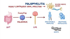
Poliomyelitis
Poliomyelitis is a highly contagious viral infection caused by the poliovirus. It enters the CNS by infecting muscle fibers and traveling retrogradely along motor neurons to the spinal cord. This can result in severe nerve injury, irreversible paralysis, breathing difficulties, and death. Other pathogens, such as herpes simplex, rabies, and tetanus, use similar retrograde axonal transport mechanisms.
Brain Fog
Microglia can reduce the number of oligodendrocyte precursors, leading to myelin loss and slower impulse conduction.
Psychiatric Disorders and Neurotransmitters
Schizophrenia: Excess dopamine; treated with dopamine receptor blockers.
Depressive Disorders: Deficiency in serotonin, norepinephrine, or dopamine; treated with SSRIs and other drugs that enhance neurotransmitter levels.
Anxiety Disorders: Abnormal norepinephrine, serotonin, and GABA activity; treated with drugs that enhance GABA or modulate norepinephrine.
Bipolar Disorder: Alternating mania and depression; treated with drugs that block sodium channels to reduce action potential generation.
Review Questions
Which PNS subdivision would a nerve injury be most life-threatening? (Somatic Motor Division)
Which cells form cerebrospinal fluid? (Ependymal Cells)
The influx of positive ions making the membrane potential less negative is called: (Depolarization)
Which is NOT a way synaptic transmission is terminated? (Reuptake into the postsynaptic neuron)
Which is an inhibitory neurotransmitter? (Glycine)
