 Back
Lecture Test 1 Review: Blood, Cardiovascular, Lymphatic, and Immune Systems
Study Guide - Smart Notes
 Tailored notes based on your materials, expanded with key definitions, examples, and context.
Blood (Chapter 17)
Composition, Physical Characteristics, and Functions
Blood is a specialized connective tissue with multiple functions essential for homeostasis. It consists of plasma (the liquid matrix) and formed elements (cells and cell fragments).
Plasma: Mostly water, contains proteins (albumin, globulins, fibrinogen), nutrients, hormones, and waste products.
Formed Elements: Erythrocytes (RBCs), leukocytes (WBCs), and platelets.
Functions: Transport of gases, nutrients, and wastes; regulation of pH, temperature, and fluid volume; protection against blood loss and infection.
Hematopoiesis
Hematopoiesis is the process of blood cell formation, occurring primarily in the red bone marrow.
All blood cells derive from hematopoietic stem cells.
Anemia Types
Anemia is a condition characterized by a deficiency of RBCs or hemoglobin, leading to reduced oxygen-carrying capacity.
Types: Blood loss anemia, iron-deficiency anemia, pernicious anemia, sickle cell anemia, and others.
Blood Typing: ABO and Rh Systems
Blood types are determined by the presence or absence of specific antigens on RBC membranes.
ABO Groups: Type A (A antigen, anti-B antibody), Type B (B antigen, anti-A antibody), Type AB (A and B antigens, no antibodies), Type O (no antigens, both anti-A and anti-B antibodies).
Rh System: Rh+ (D antigen present), Rh- (D antigen absent). Important in hemolytic disease of the newborn.
Hemolytic Disease of the Newborn
Occurs when an Rh- mother carries an Rh+ fetus, leading to maternal antibody production against fetal RBCs in subsequent pregnancies.
Transfusion Reactions
Result from mismatched blood transfusions, causing agglutination and hemolysis of donor RBCs.
Formed Elements
Erythrocytes (RBCs): Biconcave, anucleate cells specialized for oxygen transport via hemoglobin.
Hemoglobin: Protein with four polypeptide chains, each containing a heme group that binds oxygen. Broken down in the spleen and liver.
Erythropoiesis: RBC production regulated by erythropoietin (EPO), primarily from the kidneys in response to hypoxia.
Leukocytes (WBCs): Immune cells classified as granulocytes (neutrophils, eosinophils, basophils) and agranulocytes (lymphocytes, monocytes).
Platelets: Cell fragments essential for blood clotting (hemostasis).
Hemostasis
Hemostasis is the process of stopping bleeding, involving three steps:
Vascular Spasm: Vasoconstriction reduces blood flow.
Platelet Plug Formation: Platelets adhere to exposed collagen and aggregate.
Coagulation: Thrombin converts soluble fibrinogen to insoluble fibrin, forming a stable clot.
Clot Retraction: Platelets contract, pulling wound edges together and expelling serum.
PDGF and VEGF: Promote repair of smooth muscle and endothelial cells, respectively.
Fibrinolysis: Plasmin digests fibrin, removing the clot after healing.
Thrombus: Stationary clot; Embolus: Traveling clot; Embolism: Embolus lodged in a vessel, causing blockage.
The Heart (Chapter 18)
Heart Anatomy and Blood Flow
The heart is a four-chambered organ with two atria and two ventricles. Blood flows through the heart in a specific sequence, passing through valves that ensure unidirectional flow.
Valves: Atrioventricular (AV) valves (tricuspid and mitral) and semilunar (SL) valves (pulmonary and aortic).
Pulmonary Circuit: Right side pumps deoxygenated blood to the lungs.
Systemic Circuit: Left side pumps oxygenated blood to the body.
Cardiac Muscle and Conduction System
Cardiac muscle cells are striated, branched, and interconnected by intercalated discs, allowing the heart to function as a syncytium. Cardiac muscle differs from skeletal muscle in its automaticity and longer refractory period.
Intrinsic Conduction System: Specialized autorhythmic cells generate and conduct impulses, coordinating contraction.
Pacemaker: The sinoatrial (SA) node initiates the heartbeat.
AV Node Delay: Allows atria to contract before ventricles.
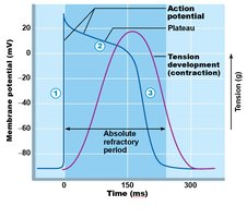
Cardiac Muscle Action Potential
Cardiac muscle action potentials involve sodium, calcium, and potassium ions, resulting in a prolonged plateau phase and refractory period.
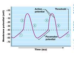
Autorhythmic Cell Action Potential
Autorhythmic cells exhibit pacemaker potentials due to spontaneous depolarization, leading to action potentials that trigger heartbeats.
Cardiac Cycle and Output
The cardiac cycle consists of alternating periods of contraction (systole) and relaxation (diastole):
Ventricular Filling: Blood flows from atria to ventricles; atrial contraction tops off ventricular volume.
Isovolumetric Contraction: Ventricles contract with all valves closed, increasing pressure.
Ventricular Ejection: SL valves open, blood is ejected.
Isovolumetric Relaxation: Ventricles relax, all valves closed.
Cardiac Output (CO): Volume of blood pumped per minute.
Stroke Volume (SV): Volume of blood pumped per beat.
End-Diastolic Volume (EDV): Volume in ventricle at end of filling.
End-Systolic Volume (ESV): Volume remaining after contraction.
Venous Return: Amount of blood returning to the heart; influences EDV and SV.
Blood Vessels (Chapter 19)
Structure of Blood Vessels
Blood vessels have three layers (tunics):
Tunica Intima: Endothelial lining; reduces friction.
Tunica Media: Smooth muscle and elastin; controls vessel diameter.
Tunica Externa: Connective tissue; protects and anchors vessels.
Types and Functions of Vessels
Arteries: Carry blood away from the heart; elastic arteries buffer pressure, arterioles regulate flow to capillaries.
Capillaries: Site of exchange; types include continuous (least permeable), fenestrated (more permeable), and sinusoid (most permeable).
Precapillary Sphincters: Regulate blood flow into capillary beds.
Veins: Carry blood toward the heart; act as blood reservoirs, have valves to prevent backflow.
Blood Pressure Regulation
Blood Pressure (BP): Force exerted by blood on vessel walls; influenced by cardiac output (CO), peripheral resistance (PR), and blood volume.
Short-Term Regulation: Neural (baroreceptors, vasomotor center) and hormonal (angiotensin II, epinephrine, norepinephrine, ADH increase BP; ANP decreases BP).
Long-Term Regulation: Renal mechanisms adjust blood volume directly (urine output) or indirectly (RAAS system).
Autoregulation: Local control of blood flow based on tissue needs (metabolic and myogenic mechanisms).
Special Circulations
Skeletal Muscle, Brain, Skin: Blood flow regulated according to activity and temperature.
Pulmonary Circulation: Low pressure; optimizes gas exchange.
Lymphatic System (Chapter 20)
Lymph and Lymphatic Vessels
Lymph is interstitial fluid collected by lymphatic capillaries and returned to the bloodstream.
Lymphatic Capillaries: More permeable than blood capillaries; absorb proteins and debris.
Right Lymphatic Duct: Drains right upper body; Thoracic Duct: Drains rest of body.
Venous Return: Ducts empty into subclavian veins.
Mechanisms: Skeletal muscle pump, respiratory pump, and valves aid lymph return.
Lymphoid Organs and Tissues
Lymph Nodes: Filter lymph, activate immune response; fewer efferent than afferent vessels slow flow for filtration.
Spleen: Filters blood, recycles RBCs, stores platelets.
Thymus: Site of T cell maturation.
MALT (Mucosa-Associated Lymphoid Tissue): Found in mucous membranes; protects against pathogens.
Tonsils: Trap and remove pathogens entering through the mouth/nose.
Peyer’s Patches: In small intestine; monitor intestinal bacteria and prevent pathogen growth.
Immune System (Chapter 21)
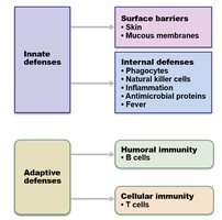
Lines of Defense
The immune system protects against pathogens through innate (nonspecific) and adaptive (specific) defenses.
First Line: Physical barriers (skin, mucous membranes).
Second Line: Internal defenses (phagocytes, natural killer cells, inflammation, antimicrobial proteins, fever).
Third Line: Adaptive immunity (B and T lymphocytes).
Inflammation and Phagocytes
Inflammation: Characterized by redness, heat, swelling, and pain; histamine is a key mediator.
Stages: Vasodilation, increased permeability, phagocyte mobilization.
First Phagocyte: Neutrophils; Second: Monocytes (become macrophages).
Antimicrobial Proteins and Complement
Interferons: Protect cells from viral infection.
Complement: Enhances phagocytosis, inflammation, and cell lysis.
Antigens and MHC
Antigen: Substance that triggers an immune response; Epitope: Specific region recognized by lymphocytes.
MHC I: Present on all nucleated cells; display endogenous antigens.
MHC II: Present on antigen-presenting cells; display exogenous antigens.
Lymphocytes: Types and Functions
B Cells: Humoral immunity; mature in bone marrow, produce antibodies after activation and clonal selection. Most become plasma cells, some become memory cells.
T Cells: Cellular immunity; mature in thymus. Types include cytotoxic (Tc), helper (Th), and regulatory (Treg) cells.
Tc Cells: Kill infected or abnormal cells; require Th cell activation.
Memory Cells: Provide faster, stronger response upon re-exposure.
Immunity Types and Disorders
Active Immunity: Natural (infection) or artificial (vaccination).
Passive Immunity: Natural (maternal antibodies) or artificial (injection of antibodies).
Vaccines: Stimulate adaptive immunity without causing disease.
AIDS: Caused by HIV; impairs immune function.
Autoimmune Disease: Immune system attacks self; treated with immunosuppressants.
Aging: Immune function declines with age.
