 Back
BackLymphatic and Respiratory Systems: Structure and Function
Study Guide - Smart Notes
 Tailored notes based on your materials, expanded with key definitions, examples, and context.
Lymphatic System
Circulatory vs. Cardiovascular vs. Lymphatic Vessels
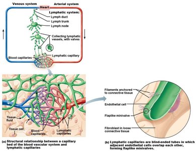
The human body contains several types of vessels that transport fluids. Understanding the distinctions between these vessels is essential for grasping the structure and function of the lymphatic system.
Circulatory vessels: A generic term that can refer to both cardiovascular and lymphatic vessels.
Cardiovascular vessels: These vessels (arteries, veins, capillaries) carry blood throughout the body.
Lymphatic vessels: These vessels transport lymph, a fluid derived from interstitial fluid. Lymphatic capillaries are blind-ended and have overlapping endothelial cells that form one-way minivalves. Lymphatic vessels generally lack tunics but contain valves to prevent backflow.
Lymph Nodes vs. Lymph Nodules
Lymph nodes and lymph nodules are both lymphatic tissues but differ in structure and location.
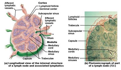
Lymph nodes: Encapsulated structures located along lymphatic vessels. They filter lymph before it returns to the venous system, have multiple afferent and fewer efferent vessels, and possess a capsule.
Lymph nodules: Unencapsulated clusters of lymphoid tissue found within the submucosal layer of mucous membranes (MALT).
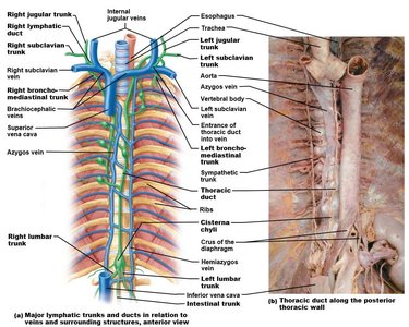
Lymph Drainage and Major Lymphatic Vessels
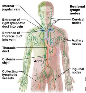
Lymph is collected from tissues and returned to the bloodstream via a network of vessels and ducts. Lymph nodes are clustered in specific regions, and lymphatic trunks drain into major ducts.
Superficial clusters of lymph nodes: Cervical, axillary, and inguinal regions.
Deep clusters of lymph nodes: Tracheobronchial, aortic, and iliac regions.
Lymphatic ducts:
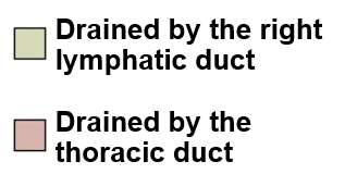
Thoracic duct: Drains the entire left half of the body, right pelvis, and right lower limb.
Right lymphatic duct: Drains the right upper quadrant of the body.
Lymphatic trunks: Jugular, subclavian, bronchomediastinal, intestinal (unpaired), and lumbar trunks collect lymph from various regions and drain into the ducts.
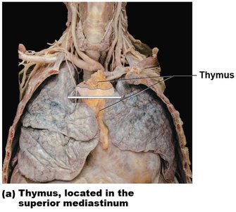
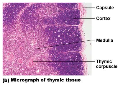
Lymphoid Organs: Thymus
The thymus is a primary lymphoid organ essential for T-cell maturation. T-lymphocytes are produced in red bone marrow and migrate to the thymus, where they acquire cell differentiation markers. The thymus is organized into lobules, each with a cortex and medulla, and contains thymic corpuscles in the medulla.
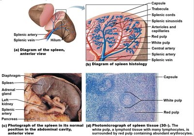
Lymphoid Organs: Spleen
The spleen is an unpaired organ located in the upper left quadrant, posterior to the stomach. It is surrounded by a capsule with inward extensions called trabeculae. The spleen contains two main tissue types:
Red pulp: Rich in blood supply, contains splenic sinusoids and cords.
White pulp: Lymphoid tissue involved in immune responses.
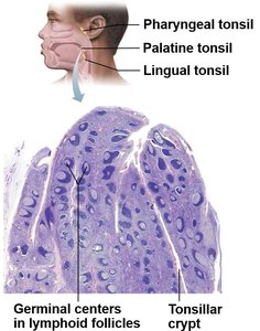
Lymphoid Organs: Tonsils
Tonsils are collections of lymphoid tissue located in the pharynx. They are part of the mucosa-associated lymphoid tissue (MALT) and are characterized by the presence of crypts surrounded by lymphoid follicles.
Palatine tonsils: Located on the lateral sides of the pharynx.
Lingual tonsil: Located at the posterior tongue.
Pharyngeal tonsil: Located on the roof of the pharynx.
Tubal tonsils: Located at the opening of the pharyngotympanic tubes.
Respiratory System
Respiratory Tracts: Upper (Conducting) vs. Lower (Respiratory) Zones
The respiratory system is divided into conducting and respiratory zones. The primary function is ventilation and external respiration.
Conducting zone: Includes structures that warm and moisten air but do not participate in gas exchange (anatomical dead space, ~150 mL).
Respiratory zone: Includes structures where external respiration (gas exchange) occurs.
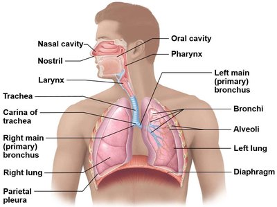
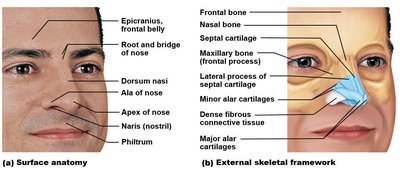
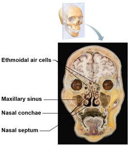
Conducting Zone: Nasal Cavity
The nasal cavity extends from the external nares to the posterior nasal apertures and is divided by the nasal septum. It is lined with mucous membranes (olfactory and respiratory mucosa) and separated from the oral cavity by the hard and soft palates.
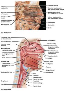
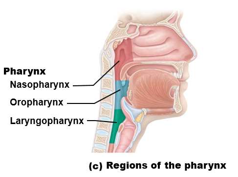
Conducting Zone: Pharynx
The pharynx is a common chamber for the respiratory and digestive tracts, extending from the base of the skull to the level of the C6 vertebra. It is divided into three regions:
Nasopharynx: Air passage only; closed off during swallowing by the uvula and soft palate.
Oropharynx: Extends from the soft palate to the epiglottis.
Laryngopharynx: Posterior to the larynx.
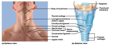
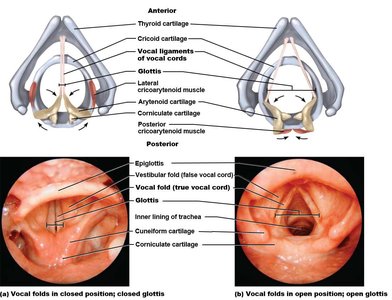
Conducting Zone: Larynx
The larynx is a passageway for air only, connecting the laryngopharynx to the trachea. It is supported by nine cartilages and is responsible for sound production.
Thyroid cartilage: Forms the laryngeal prominence (Adam's apple).
Cricoid cartilage: Complete ring, narrow anteriorly.
Arytenoid, corniculate, and cuneiform cartilages: Posterior support and anchor the vocal cords.
Epiglottis: Elastic cartilage that closes the airway during swallowing.
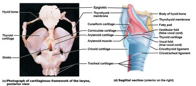
Vocal Folds and Glottis
The larynx contains two sets of folds:
Vestibular folds: "False" vocal cords, superior to the true vocal cords.
Vocal folds: "True" vocal cords, responsible for sound production via abduction and adduction during exhalation.
Glottis: The space between the vocal folds (rima glottidis).
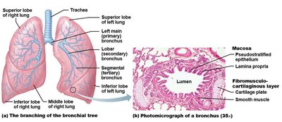
Conducting Zone: Trachea
The trachea is a flexible tube anterior to the esophagus, lined with pseudostratified ciliated columnar epithelium. It contains 16-20 C-shaped rings of hyaline cartilage, open posteriorly to allow esophageal expansion during swallowing. The trachea bifurcates at the carina (T7 vertebra) into primary bronchi.

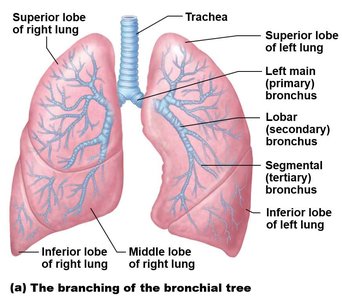
Conducting Zone: Bronchi and Bronchioles
The bronchial tree branches extensively within the lungs:
Primary bronchi: Enter each lung at the hilum.
Secondary (lobar) bronchi: Supply each lobe of the lungs.
Tertiary (segmental) bronchi: Supply bronchopulmonary segments.
Bronchioles: Smaller airways lacking cartilage, with abundant smooth muscle.
Terminal bronchioles: Last segment of the conducting zone.
Respiratory bronchioles: First segment of the respiratory zone, some smooth muscle remains.
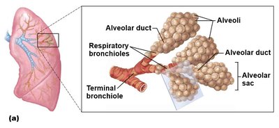
Respiratory Zone: Alveoli
The alveoli are the primary sites of gas exchange in the lungs. Alveolar ducts connect respiratory bronchioles to terminal alveoli. Alveoli are lined by two main cell types:
Type I alveolar cells: Simple squamous cells, 97% of alveolar surface, facilitate gas exchange.
Type II alveolar cells: Cuboidal cells, produce surfactant to reduce surface tension.
Thoracic Cavity: Gross Anatomy
Each lung is contained within a double-layered pleural membrane. The left lung is slightly smaller due to the cardiac notch and has two lobes (superior and inferior), while the right lung has three lobes (superior, middle, and inferior) separated by horizontal and oblique fissures.
Lung | Lobes | Fissures |
|---|---|---|
Left | 2 (Superior, Inferior) | Oblique |
Right | 3 (Superior, Middle, Inferior) | Horizontal, Oblique |
Root of the lung: The mediastinal surface where bronchi, vessels, and nerves enter and exit the lung.
