 Back
BackMembrane Transport, Membrane Potential, and Skeletal Muscle Structure: Study Notes for Human Anatomy & Physiology I
Study Guide - Smart Notes
 Tailored notes based on your materials, expanded with key definitions, examples, and context.
Exam 2 Part 1
Membrane Transport and Permeability
Cell Membrane as a Selective Barrier
The cell membrane separates the intracellular environment from the extracellular space, allowing cells to maintain distinct concentrations of electrolytes and solutes. This selective permeability is essential for cellular homeostasis.
Intracellular fluid (cytosol): Higher concentration of potassium ions (K+) and negatively charged proteins.
Extracellular fluid (interstitial): Higher concentration of sodium ions (Na+), chloride ions (Cl-), and calcium ions (Ca2+).
Permeability: The ease with which substances cross the membrane. Cell membranes are selectively permeable, allowing some substances to pass while restricting others.
Transport Mechanisms
Substances move across cell membranes via passive or active transport mechanisms:
Passive transport: No energy required; includes diffusion and facilitated diffusion.
Active transport: Requires energy (ATP); includes ion pumps and secondary active transport.
Diffusion
Diffusion is the movement of molecules from an area of high concentration to low concentration, driven by random molecular motion.
Chemical gradient: Drives diffusion of uncharged molecules.
Electrochemical gradient: Drives diffusion of charged ions, influenced by both concentration and electrical gradients.
Examples: Oxygen (O2) diffuses from lungs to blood; carbon dioxide (CO2) diffuses from cells to blood.
Osmosis
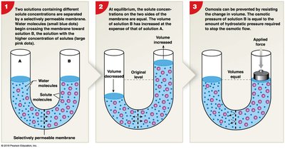
Osmosis is the diffusion of water across a semi-permeable membrane in response to solute concentration differences.
Water moves toward higher solute concentration.
Osmotic pressure: The force required to prevent water movement into a solution.
Tonicity
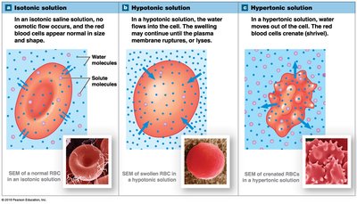
Tonicity describes the effect of extracellular solutions on cell volume:
Isotonic: No net water movement; cell volume remains unchanged.
Hypotonic: Water enters the cell; cell may swell and burst (cytolysis).
Hypertonic: Water leaves the cell; cell shrivels (crenation).
Factors Influencing Diffusion
Distance: Effective only over short distances.
Molecule size: Smaller molecules diffuse faster.
Temperature: Higher temperature increases diffusion rate.
Concentration gradient: Larger gradient increases diffusion rate.
Electrical gradient: For ions, electrical forces also influence movement.
Carrier-Mediated Transport
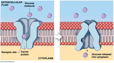
Carrier proteins in the membrane facilitate the transport of specific molecules or ions:
Specificity: Each carrier protein transports a particular substance.
Saturation limits: Maximum rate determined by number of carrier proteins.
Regulation: Activity can be controlled by various factors.
Cotransport: Two substances move in the same direction.
Counter-transport: Two substances move in opposite directions.
Facilitated Diffusion
Facilitated diffusion is a passive process where substances move down their concentration gradient with the help of a transport protein. No ATP is required.
Membrane Potential and Bioelectricity
Trans-Membrane Potential
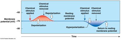
The trans-membrane potential is the difference in electrical charge across the cell membrane. At rest, cells have a more negative charge inside compared to outside.
Resting membrane potential: Typically -70 mV in nerve cells, -85 mV in muscle cells.
Electrochemical gradient: Sum of chemical and electrical forces acting on ions across the membrane.
Sodium-potassium pump: Maintains resting potential by moving Na+ out and K+ in.
Ion Channels and Gated Channels
Chemically regulated channels: Open or close in response to binding specific chemicals (e.g., neurotransmitters).
Voltage-regulated channels: Open or close in response to changes in membrane potential.
Depolarization, Hyperpolarization, and Repolarization
Depolarization: Membrane potential becomes less negative.
Hyperpolarization: Membrane potential becomes more negative.
Repolarization: Membrane potential returns toward resting value after depolarization.
Skeletal Muscle Structure and Function
Types of Muscle Tissue
Muscle tissue is specialized for contraction and exists in three forms:
Skeletal muscle: Striated, voluntary, attached to bones.
Cardiac muscle: Striated, involuntary, found in the heart.
Smooth muscle: Non-striated, involuntary, lines hollow organs.
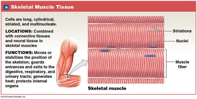
Skeletal Muscle Functions
Produce movement
Maintain posture
Support soft tissues
Guard entrances and exits
Maintain body temperature
Store nutrient reserves
Gross Anatomy of Skeletal Muscles
Origin: Attachment to stationary bone.
Insertion: Attachment to moving bone.
Synergistic muscles: Work together for a common action.
Antagonistic muscles: Oppose each other's actions (e.g., flexors vs. extensors).
Organization of Connective Tissues
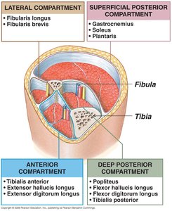
Endomysium: Covers individual muscle fibers.
Perimysium: Sheathes bundles of muscle fibers (fascicles).
Epimysium: Surrounds the entire muscle.
Deep fascia: Wraps groups of cooperating muscles.
Skeletal Muscle Cells (Muscle Fibers)
Multinucleate, very long cells formed by fusion of myoblasts.
Limited repair; new cells from satellite cells.
Structure of a Skeletal Muscle Cell
Myofilaments: Actin (thin) and myosin (thick).
Myofibrils: Bundles of myofilaments.
Sarcoplasm: Muscle cell cytoplasm.
Sarcolemma: Cell membrane, excitable.
Sarcoplasmic reticulum (SR): Modified ER, stores Ca2+.
Transverse tubules (T-tubules): Conduct action potentials deep into cell.
Sarcomere Structure
Sarcomere: Repeating unit of myofilaments in myofibril.
I band: Light band, only thin filaments.
A band: Dark band, thick filaments and overlap.
H band: Only thick filaments.
Z disk: Border between sarcomeres.
Sliding Filament Model of Muscle Contraction
Muscle contraction occurs as thin actin filaments slide over thick myosin filaments, shortening the sarcomere.
Myosin heads form cross bridges with actin, pulling actin toward the center.
ATP is required for both cross bridge formation and release.
Calcium ions released from SR bind to troponin, moving tropomyosin and exposing actin sites.
Summary Table: Muscle Cell Structures
Structure | Function |
|---|---|
Sarcolemma | Excitable membrane, conducts action potentials |
Sarcoplasmic Reticulum | Stores Ca2+ for contraction |
Myofibril | Bundle of myofilaments, contracts |
Actin (thin) | Slides over myosin, shortens sarcomere |
Myosin (thick) | Forms cross bridges, pulls actin |
Key Terms and Definitions
Electrolyte: Substance that dissociates into ions in solution, conducting electricity.
Anion: Negatively charged ion (e.g., Cl-).
Cation: Positively charged ion (e.g., Na+, K+).
Homeostasis: Maintenance of stable internal environment.
Action potential: Rapid change in membrane potential, essential for muscle and nerve function.
Sample Questions
Which of the following are anions?
A. Sodium ions and potassium ions
B. Chloride ions
C. Calcium ions
D. All of the above are anions
E. None of the above are anions
If binding acetylcholine to a chemically gated sodium ion channel opens the channel, what happens?
A. Sodium ions will move into the cell causing a hyperpolarization
B. Sodium ions will move out of the cell causing a hyperpolarization
C. Sodium ions will move into the cell causing a depolarization
D. Sodium ions will move out of the cell causing a depolarization
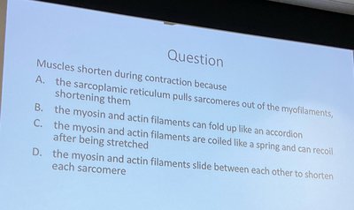
Muscles shorten during contraction because:
A. The sarcoplasmic reticulum pulls sarcomeres out of the myofilaments, shortening them
B. The myosin and actin filaments can fold up like an accordion
C. The myosin and actin filaments are coiled like a spring and can recoil after being stretched
D. The myosin and actin filaments slide between each other to shorten each sarcomere
Equations and Formulas
Osmotic Pressure: Where is osmotic pressure, is the van't Hoff factor, is molarity, is the gas constant, and is temperature.
Resting Membrane Potential: Where is membrane potential, is gas constant, is temperature, is Faraday's constant, and are potassium concentrations outside/inside.
Additional info: Academic context and expanded explanations were added to clarify fragmented notes and ensure completeness for exam preparation.
