 Back
BackRepro 1 - March 20
Study Guide - Smart Notes
 Tailored notes based on your materials, expanded with key definitions, examples, and context.
Metabolism and Its Regulation
Fed State Metabolism
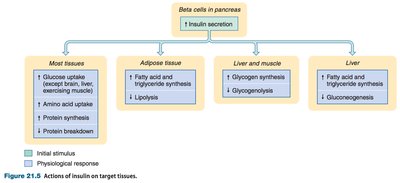
The fed state, or absorptive state, occurs after eating when nutrients are being absorbed and processed. The body focuses on storing energy and building macromolecules.
Skeletal Muscle: Uptakes glucose for energy and stores it as glycogen (about 70% of body glycogen). Amino acids are used for protein turnover.
Liver: Converts glucose to glycogen (24% of body stores) and fatty acids (which are transported to adipocytes). Amino acids are used for protein synthesis or converted to keto acids for energy or fat synthesis.
Adipocytes: Take up dietary triglycerides from chylomicrons and convert excess glucose to triglycerides. Fat storage is essentially unlimited, while glycogen and protein storage are limited.
Key Processes:
Glycogenesis: Formation of glycogen from glucose in liver and muscle.
Lipogenesis: Synthesis of fatty acids and triglycerides from glucose.
Protein Synthesis: Uptake of amino acids for building proteins, especially in muscle.
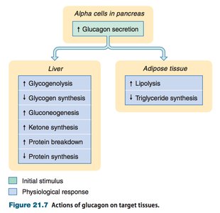
Fasted State Metabolism
The fasted state, or postabsorptive state, occurs between meals. The body relies on stored energy to maintain blood glucose, which is critical for nervous system function.
Skeletal Muscle: Breaks down glycogen to glucose-6-phosphate for its own use, producing pyruvate and lactate.
Liver: Converts glycogen to glucose (glycogenolysis) and produces new glucose from pyruvate, lactate, glycerol, and certain amino acids (gluconeogenesis). Also converts fatty acids to ketone bodies for energy.
Adipocytes: Undergo lipolysis, releasing fatty acids and glycerol into the bloodstream for energy use by most cells.
Key Processes:
Glycogenolysis: Breakdown of glycogen to glucose.
Lipolysis: Breakdown of triglycerides to fatty acids and glycerol.
Gluconeogenesis: Synthesis of new glucose from non-carbohydrate sources.
Ketogenesis: Formation of ketone bodies from fatty acids.
Protein Degradation: Breakdown of proteins to amino acids for gluconeogenesis or energy.
Regulation of Metabolism: Hormonal Control
Metabolic states are primarily regulated by the hormones insulin (fed state) and glucagon (fasted state).
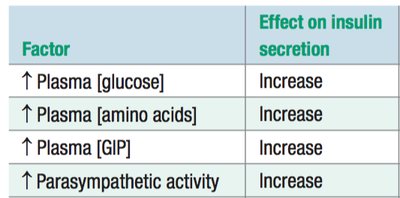
Insulin: Secreted by pancreatic beta cells in response to increased plasma glucose, amino acids, GIP, and parasympathetic activity. Promotes anabolism (energy storage and synthesis of macromolecules).
Glucagon: Secreted by pancreatic alpha cells in response to decreased plasma glucose and sympathetic activity. Promotes catabolism (breakdown of energy stores).
Factor | Effect on Insulin Secretion |
|---|---|
↑ Plasma [glucose] | Increase |
↑ Plasma [amino acids] | Increase |
↑ Plasma [GIP] | Increase |
↑ Parasympathetic activity | Increase |
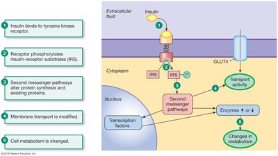
Cellular Mechanisms of Insulin Action
Insulin acts via a tyrosine kinase receptor, leading to a cascade of intracellular events that increase glucose uptake and alter metabolism.
Binding of insulin to its receptor triggers phosphorylation of insulin-receptor substrates (IRS).
Second messenger pathways activate protein synthesis and modify existing proteins.
Membrane transport is modified, notably by insertion of GLUT4 glucose transporters into the cell membrane.
Overall, cell metabolism is shifted toward storage and synthesis.
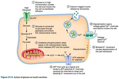
Cellular Mechanisms of Glucose-Stimulated Insulin Secretion
Glucose enters beta cells via GLUT2 transporters, is metabolized to produce ATP, which closes ATP-sensitive K+ channels, depolarizing the cell and opening voltage-gated Ca2+ channels. The resulting Ca2+ influx triggers insulin exocytosis.
Reproduction and Development
Sex Determination and Chromosomes
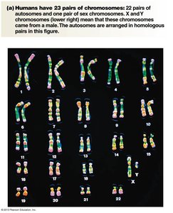
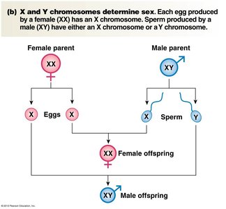
Sex determination is programmed in the genome. Each nucleated cell (except gametes) contains 23 pairs of chromosomes: 22 pairs of autosomes and 1 pair of sex chromosomes (XX for females, XY for males). Gametes are haploid, containing 23 single chromosomes.
Autosomes: Direct general body development.
Sex Chromosomes: Direct development of internal and external sex organs.
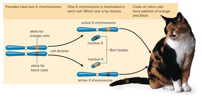
X-Chromosome Inactivation
In females, one X chromosome is randomly inactivated in each cell during early embryonic development. This process ensures dosage compensation between males and females and explains why X-linked recessive disorders are more common in males.
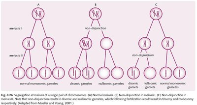
Abnormal Sex Chromosome Distribution
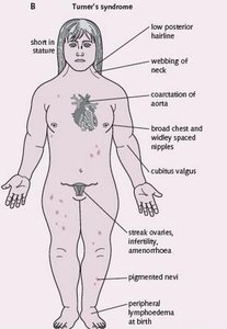
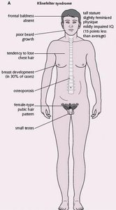
Errors in chromosome segregation (nondisjunction) during meiosis can lead to abnormal numbers of sex chromosomes, resulting in syndromes such as Turner (XO) and Klinefelter (XXY).
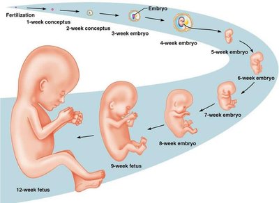
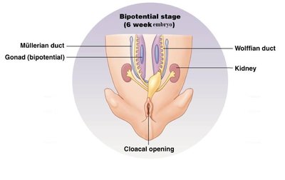
Embryonic Development and Sex Differentiation
The embryonic period extends through the 8th week. Early in development, reproductive structures are bipotential and can develop into either male or female organs depending on genetic and hormonal signals.
Genetic and Hormonal Control of Sex Differentiation
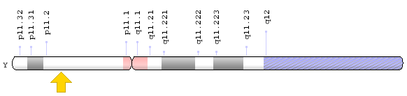
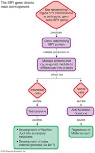
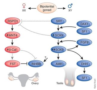
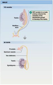
The presence or absence of the SRY gene on the Y chromosome determines gonadal differentiation. The SRY gene produces testis-determining factor (TDF), which initiates testis development. Testes then secrete hormones that drive male differentiation:
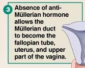
Anti-Müllerian Hormone (AMH): Causes regression of Müllerian ducts (precursors to female internal structures).
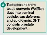
Testosterone: Converts Wolffian ducts into male accessory structures (epididymis, vas deferens, seminal vesicles).
Dihydrotestosterone (DHT): Drives differentiation of external male genitalia.
If Female | If Male |
|---|---|
Gonad (cortex) forms ovary Gonad (medulla) regresses Wolffian duct regresses (testosterone absent) Müllerian duct becomes fallopian tube, uterus, cervix, and upper 1/2 of vagina (AMH absent) | Gonad (cortex) regresses Gonad (medulla) forms testis Wolffian duct forms epididymis, vas deferens, and seminal vesicle (testosterone present) Müllerian duct regresses (AMH present) |
Example: In the absence of the SRY gene, the gonads develop into ovaries, and the Müllerian ducts form the female reproductive tract. In the presence of SRY, testes develop and secrete hormones that drive male differentiation.
Additional info: Disorders of sexual development can arise from mutations in the SRY gene, androgen insensitivity, or abnormal hormone production, leading to atypical differentiation of internal or external genitalia.
