 Back
BackMicroscopy: Structure, Function, and Application in Anatomy & Physiology
Study Guide - Smart Notes
 Tailored notes based on your materials, expanded with key definitions, examples, and context.
The Microscope in Anatomy & Physiology
Introduction to Microscopy
The microscope is an essential tool in anatomy and physiology, allowing for the visualization of structures not visible to the naked eye. Understanding its parts, functions, and proper usage is foundational for laboratory work in biological sciences.
Parts and Functions of the Microscope
Major Components
Ocular (Eyepiece): The lens you look through, typically with 10X magnification.
Revolving Nosepiece: Holds and allows rotation of objective lenses.
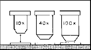
Objective Lenses: Provide different magnification powers (commonly 4X, 10X, 40X, 100X).
Stage: Platform where the slide is placed.
Stage Motion Knobs: Move the slide horizontally and vertically.
Light Source: Illuminates the specimen.
Coarse Adjustment Knob: Moves the stage up and down for general focusing.
Fine Adjustment Knob: Allows for precise focusing.
Power Switch/Brightness Control: Turns the microscope on/off and adjusts light intensity.
Condenser Lever: Adjusts the amount of light reaching the specimen.
Stage Clips: Hold the slide in place.
Iris Diaphragm Lever: Controls the diameter of the light beam (cone of light) passing through the condenser.
Condenser: Focuses light onto the specimen.
Microscope Terminology
Key Terms and Definitions
Resolution: The ability to distinguish two points as separate entities. Higher resolution reveals more detail.
Parfocal: Property that allows the microscope to stay in focus when switching between objective lenses.
Working Distance: The distance between the objective lens and the specimen when in focus. Higher magnification lenses have shorter working distances.
Field of Vision (Field of View): The visible area seen through the microscope at a given magnification.
Depth of Field: The thickness of the specimen that remains in focus at one time.
Magnification and Calculations
Total Magnification
Total magnification is calculated by multiplying the magnification of the ocular lens by that of the objective lens:
Example: If the ocular is 10X and the objective is 40X, total magnification is .
Field of Vision Calculations
The field of view decreases as magnification increases. If you know the field of view at one magnification, you can calculate it for another using:
Example: If FOV at 4X is 4.5 mm, then at 40X: so .
Estimating Object Size
Estimate the size of an object by comparing it to the known field of view. For example, if a cell spans half the field at 0.45 mm, its size is approximately 0.225 mm.
Microscope Operation and Techniques
Proper Focusing Technique
Start with the lowest power objective (usually 4X).
Use the coarse adjustment knob to bring the specimen into general focus.
Switch to higher power objectives as needed, using only the fine adjustment knob for focusing.
Microscopes are parfocal, so only minor adjustments are needed when changing objectives.
Orientation of Specimens
Images appear upside down and reversed under the microscope due to the optics.

Field of Vision and Depth of Field
Field of Vision
As magnification increases, the field of vision decreases.
Field of vision can be measured using a grid slide with known dimensions.
Depth of Field
Depth of field is the vertical range that remains in focus at one time.
Higher magnification lenses have a shallower depth of field.
Wet Mount Preparation
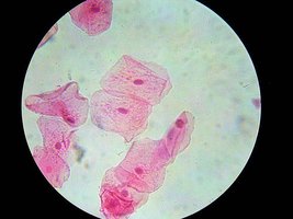
Wet Mount of Cheek Cells
Wet mounts are prepared by placing a drop of liquid (e.g., water or stain) on the specimen and covering it with a cover slip.
This technique is commonly used for viewing cells, such as human cheek cells.

Sample Questions and Applications
Which part controls the cone of light? The iris diaphragm lever.
If FOV at 4X is 4.5 mm, what is FOV at 40X? 0.45 mm.
Total magnification at 40X objective? 400X (assuming 10X ocular).
True or False: Working distance at 4X is less than at 10X? False; working distance decreases as magnification increases.
Focusing at 10X should start with fine adjustment? False; start with coarse adjustment at low power, then fine adjustment at higher powers.
Are images upside down and backwards? True; due to the microscope's optics.
Summary Table: Microscope Lenses and Field of View
Objective Lens | Magnification | Field of View (mm) | Working Distance |
|---|---|---|---|
4X | 40X (with 10X ocular) | 4.5 | Longest |
10X | 100X | 1.8 | Shorter |
40X | 400X | 0.45 | Short |
100X | 1000X | 0.18 | Shortest |
Additional info: The above table summarizes the relationship between objective lens magnification, total magnification, field of view, and working distance. As magnification increases, field of view and working distance decrease.
