 Back
Mini-Textbook Study Guide: Tissue Types and Histology in the Human Body
Study Guide - Smart Notes
 Tailored notes based on your materials, expanded with key definitions, examples, and context.
4 Types of Tissue in the Human Body
Overview of Tissue Types
The human body is composed of four primary tissue types, each with distinct functions and characteristics. Understanding these tissues is fundamental to histology, the study of normal, healthy body tissues.
Epithelial Tissue: Covers body surfaces and lines internal cavities; forms skin and linings.
Connective Tissue: Supports, binds, and protects; includes bone, tendons, fat, blood, and soft tissues.
Muscle Tissue: Responsible for movement; includes skeletal, cardiac, and smooth muscle.
Nervous Tissue: Regulates and controls body functions; includes brain, spinal cord, and nerves.
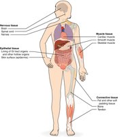
Histology and Pathohistology
Histology is the study of tissues in their normal, healthy state. When tissues are diseased, the study is referred to as pathohistology.
Embryonic Origins of Tissue Types
Germ Layers and Tissue Differentiation
During embryonic development, tissues arise from three primary germ layers: ectoderm, mesoderm, and endoderm. Each tissue type originates from specific layers:
Epithelial Tissue: Derived from all three germ layers.
Connective Tissue: Derived from mesoderm.
Muscle Tissue: Derived from mesoderm.
Nervous Tissue: Derived from ectoderm.
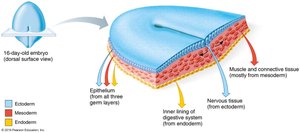
Epithelial Tissue
Functions and Forms
Epithelial tissue forms boundaries, protects, secretes, absorbs, and filters. It exists in two main forms:
Covering and lining epithelium: Skin, linings of digestive and respiratory tracts.
Glandular epithelium: Forms glands of the endocrine system.
Structural Characteristics
Epithelial tissue exhibits polarity, with two distinct surfaces:
Apical surface: Exposed to the body exterior or internal cavity; may have cilia or microvilli.
Basal surface: Attached to underlying connective tissue via the basal lamina, a thin sheet of glycoproteins and collagen fibers.
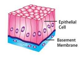
Naming Epithelial Tissue
Epithelial tissues are classified by the number of layers and cell shape:
Layers: Simple (single layer) – absorption, filtration, secretion Stratified (multiple layers) – protection
Shape: Squamous – flat, scale-like Cuboidal – cube-shaped Columnar – tall, column-like
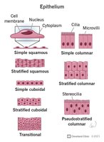
Types of Epithelial Tissue
Each form of epithelial tissue performs specialized functions:
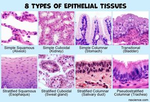
Simple Squamous: Diffusion and filtration; found in lungs, blood vessels, kidney glomeruli.
Simple Cuboidal: Secretion and absorption; found in kidney tubules.
Simple Columnar: Absorption and secretion; lines GI tract.
Pseudostratified Columnar: Absorption and secretion; lines upper respiratory tract.
Stratified Squamous: Protection; found in epidermis, mouth, esophagus, vagina.
Stratified Cuboidal: Secretion; rare, found in sweat and mammary glands.
Stratified Columnar: Secretion; rare, found in salivary glands.
Transitional Epithelium: Allows stretch; found in urinary system.
Glandular Epithelium
Exocrine vs. Endocrine Glands
Glands are classified by their mode and location of secretion:
Exocrine glands: Release products onto body surfaces or into cavities via ducts; can be unicellular (e.g., goblet cells) or multicellular.
Endocrine glands: Release hormones into the bloodstream; lack ducts and are surrounded by blood vessels.
Modes of Secretion
Merocrine: Secrete products via exocytosis; cells remain intact (e.g., salivary glands).
Holocrine: Cells rupture to release products (e.g., sebaceous glands).
Apocrine: Cells lose small portions during secretion (e.g., mammary glands).
Connective Tissue
Functions and Classes
Connective tissue is the most abundant tissue in the body, serving to bind, support, protect, insulate, and transport substances. All connective tissue originates from embryonic mesenchyme (mesoderm).
Connective tissue proper
Cartilage
Bone
Blood
Structural Components
All connective tissues share three main components:
Ground Substance (Extracellular Matrix): Unstructured material filling space between cells; contains interstitial fluid, cell adhesion proteins, and proteoglycans.
Fibers: Provide support; three types—collagen (strongest), elastic (stretchy), reticular (delicate network).
Cells: Vary by tissue type (e.g., fibroblasts in proper, chondrocytes in cartilage, osteocytes in bone, blood cells in blood).
Types of Connective Tissue Proper
Loose Areolar: Gel-like, all fiber types; wraps/cushions organs.
Loose Adipose: Stores fat; insulates and protects organs.
Loose Reticular: Network of reticular fibers; supports lymphoid organs.
Dense Regular: Parallel collagen fibers; resists tension in one direction (tendons, ligaments).
Dense Irregular: Irregular collagen fibers; withstands tension in multiple directions (dermis, organ capsules).
Dense Elastic: Many elastic fibers; allows tissue recoil (arteries, bronchial tubes).
Cartilage
Cartilage is resilient, able to withstand tension and compression, and lacks blood vessels and nerves. Types include:
Hyaline Cartilage: Supports and cushions; found in joints, ribs, nose, trachea.
Elastic Cartilage: Flexible; found in external ear and epiglottis.
Fibrocartilage: Absorbs shock; found in intervertebral discs, knee menisci.
Bone
Bone provides structural support and protection, stores minerals and fat, and is the site of blood cell formation. It consists of a hard, calcified matrix with collagen fibers and calcium salts. Bone structure includes:
Compact (Cortical) Bone: Dense, strong; contains osteons (concentric rings).
Spongy (Cancellous) Bone: Less dense, contains marrow.
Blood
Blood is an atypical connective tissue, originating from mesenchyme. It serves as the transport vehicle of the body, with plasma as its extracellular matrix and soluble protein fibers for clotting.
Muscle Tissue
Types and Functions
Muscle tissue is responsible for movement and consists of tightly packed cells with abundant blood supply. Three types are:
Skeletal Muscle: Voluntary, striated, multinucleated; attached to bones.
Cardiac Muscle: Involuntary, striated, branching; found in heart walls, connected by intercalated discs.
Smooth Muscle: Involuntary, non-striated, spindle-shaped; found in walls of hollow organs.
Nervous Tissue
Structure and Function
Nervous tissue forms the nervous system, regulating and controlling body functions. It consists of:
Neurons: Specialized cells transmitting electrochemical signals; have axons (transmitters) and dendrites (receivers).
Neuroglia: Supporting cells that insulate and protect neurons.
Tissue Protection and Healing
First-Line Protection
The body uses various structures and processes for protection:
Skin and mucosa: Mechanical boundary.
Ciliated and mucous cells: Mechanical and chemical barrier in respiratory tract.
Stomach acid: Chemical barrier.
Healing Process
When injured, tissues undergo inflammation, organization, regeneration, and fibrosis:
Inflammation: Release of chemicals, increased vessel permeability, migration of WBCs and proteins.
Organization: Restoration of blood supply, formation of granulation tissue.
Regeneration and Fibrosis: Replacement of damaged tissue, formation of scar tissue.
Regenerative Capacity of Tissues
Tissue types vary in their ability to regenerate:
Epithelial tissue, bone, and some connective tissues: Regenerate easily.
Smooth muscle and dense regular connective tissue: Moderate regeneration.
Cardiac muscle and nervous tissue: Virtually no functional regenerative capacity.
Tissue Type | Regenerative Capacity |
|---|---|
Epithelial Tissue | High |
Bone (Compact & Spongy) | High |
Connective Tissue (some forms) | High |
Smooth Muscle | Moderate |
Dense Regular Connective Tissue | Moderate |
Cardiac Muscle | Low/None |
Nervous Tissue | Low/None |
Additional info: This study guide covers content relevant to ANP college courses, specifically Chapter 4: Tissue: The Living Fabric, and provides foundational context for other chapters related to tissue structure and function.
