 Back
BackMini-Textbook Study Notes: Bones and Skeletal Tissue
Study Guide - Smart Notes
 Tailored notes based on your materials, expanded with key definitions, examples, and context.
Gross Anatomy of Bone
Overview of Bone Structure
The human skeleton is composed of bones that provide structural support, protection, and facilitate movement. Bones are complex organs containing various tissues, including bone tissue, cartilage, nerves, blood vessels, and connective tissue.
Skeletal Cartilage
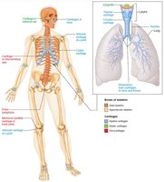
Characteristics and Types
Skeletal cartilage is avascular, not innervated, and resilient. It is surrounded by a dense irregular connective tissue called the perichondrium, which supplies oxygen and nutrients and prevents outward expansion during compression.
Hyaline Cartilage: Provides support, flexibility, and resilience; most abundant type.
Elastic Cartilage: Contains more elastic fibers; found in the external ear and epiglottis.
Fibrocartilage: Highly compressible; found in intervertebral discs and knee menisci.
Growth of Cartilage
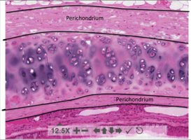
Appositional and Interstitial Growth
Cartilage grows by two mechanisms:
Appositional Growth: Chondroblasts in the perichondrium secrete new matrix on the external surface.
Interstitial Growth: Chondrocytes within lacunae divide and secrete new matrix, expanding cartilage from within.
From Cartilage to Bone
Developmental Transition
The human skeleton initially consists of cartilage and membranes, which are gradually replaced by bone during development. Calcification of cartilage occurs when calcium salts are deposited, hardening the tissue. However, hardened cartilage is not the same as bone.
Functions of Bone
Major Roles in the Body
Bones perform several essential functions:
Support: Framework for the body and soft organs.
Protection: Shields the brain, spinal cord, and vital organs.
Anchorage: Acts as levers for muscle action.
Mineral and Growth Factor Storage: Reservoir for calcium, phosphorus, and growth factors.
Blood Cell Formation: Hematopoiesis occurs in red marrow cavities.
Triglyceride Storage: Fat stored in bone cavities for energy.
Hormone Production: Osteocalcin regulates insulin secretion, glucose levels, and metabolism.
Classification of Bones
Axial vs. Appendicular Skeleton
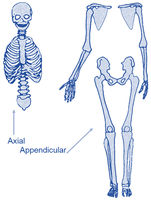
The 206 named bones are divided into two groups:
Axial Skeleton: Skull, vertebral column, rib cage.
Appendicular Skeleton: Limbs and girdles.
Bone Shapes
Bones are classified by shape:
Long Bones: Longer than wide; limb bones.
Short Bones: Cube-shaped; wrist and ankle bones, sesamoid bones (patella).
Flat Bones: Thin, flat, slightly curved; sternum, scapulae, ribs, skull bones.
Irregular Bones: Complex shapes; vertebrae, hip bones.
Compact and Spongy Bone
Bone Tissue Types
Bones contain two types of tissue:
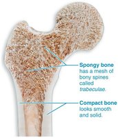
Compact Bone: Dense, smooth, and solid; provides strength.
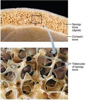
Spongy Bone: Mesh of bony spines (trabeculae); open spaces filled with marrow.
Structure of Short, Irregular, and Flat Bones
Internal and External Features
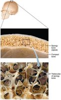
These bones have thin plates of spongy bone (diploe) covered by compact bone. The periosteum covers the outside, and the endosteum covers the inside. Bone marrow is scattered throughout spongy bone, and hyaline cartilage covers areas involved in joints.
Structure of Long Bones
Diaphysis, Epiphyses, and Membranes
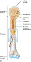
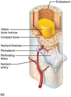
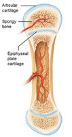
Long bones consist of a diaphysis (shaft), epiphyses (ends), and membranes. The diaphysis is a tubular shaft with compact bone surrounding the medullary cavity. Epiphyses have compact bone externally and spongy bone internally, with articular cartilage covering joint surfaces. The epiphyseal line marks the site of bone growth.
Periosteum and Endosteum
Bone Membranes
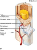
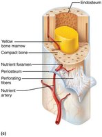
The periosteum is a double-layered membrane covering external bone surfaces except joints. It has a fibrous layer (dense irregular connective tissue) and an osteogenic layer (stem cells). The endosteum is a delicate membrane covering internal bone surfaces, including trabeculae and canals, and contains osteogenic stem cells.
Hematopoietic Tissue
Red and Yellow Marrow
Red marrow is found in spongy bone cavities and medullary cavities of newborns, most active in flat and some irregular bones. Yellow marrow is found in adult medullary cavities, producing fat, cartilage, and bone, and can convert to red marrow if needed.
Microanatomy of Bone
Microscopic Structure
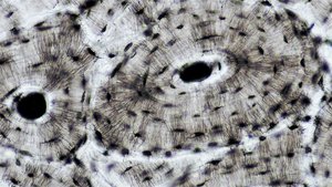
Bones are composed of specialized cells and matrix arranged in structural units called osteons. The matrix contains collagen fibers and mineral deposits, providing strength and flexibility.
Cells of Bone Tissue
Five Cell Types
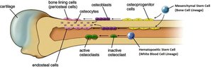
Bone tissue contains five main cell types:
Osteogenic cells: Stem cells that differentiate into osteoblasts.
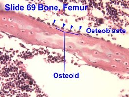
Osteoblasts: Bone-forming cells that secrete osteoid.
Osteocytes: Mature bone cells that maintain bone matrix.
Bone lining cells: Flat cells on bone surfaces, help maintain matrix.
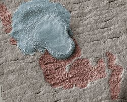
Osteoclasts: Bone-resorbing cells that break down bone matrix.
Osteons
Structural Unit of Compact Bone
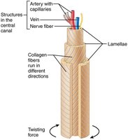
Osteons are elongated cylinders running parallel to the bone's long axis, acting as weight-bearing pillars. They consist of concentric rings of bone matrix called lamellae, with collagen fibers running in different directions in adjacent rings.
Non-Osteon Lamellae
Interstitial and Circumferential Lamellae
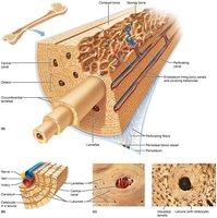
Interstitial lamellae fill gaps between forming osteons, while circumferential lamellae extend around the diaphysis, helping resist twisting forces.
Bone Canals
Central and Perforating Canals
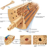
The central (Haversian) canal runs through the core of the osteon, containing blood vessels and nerves. Perforating (Volkmann's) canals run at right angles, connecting blood vessels and nerves of the periosteum, medullary cavity, and central canal. Both are lined with endosteum.
Lacunae & Canaliculi
Cellular Communication
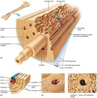
Lacunae are small cavities at the junctions of lamellae containing osteocytes. Canaliculi connect lacunae, allowing nutrients, wastes, and signals to be relayed between cells.
Spongy Bone
Structure and Function
Spongy bone appears disorganized but is structured along lines of stress. Trabeculae confer strength, contain irregularly arranged lamellae and osteocytes, and are supplied by capillaries in the endosteum.
Organic Components of Bone
Osteoid and Collagen
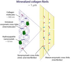
Osteoid is secreted by osteoblasts and consists of ground substance and collagen fibers, making up one-third of the organic bone matrix. Collagen provides tensile strength and flexibility, with sacrificial bonds preventing fractures.
Inorganic Components of Bone
Hydroxyapatites
Hydroxyapatites (mineral salts) make up 65% of bone mass, mainly as tiny calcium phosphate crystals around collagen fibers. They are responsible for bone hardness and resistance to compression.
Bone Growth & Development
Ossification (Osteogenesis)
Ossification is the process of bone tissue formation, beginning in the second month of fetal development and continuing postnatally until early adulthood. Bone remodeling and repair are lifelong processes.
Formation of Bony Skeleton
Types of Ossification
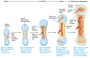
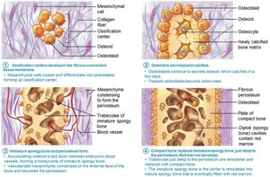
Up to week 8 of fetal development, the skeleton is fibrous membranes and hyaline cartilage. Two types of ossification occur:
Endochondral Ossification: Bone forms by replacing hyaline cartilage; forms most bones below the skull except the clavicle.
Intramembranous Ossification: Bone develops from fibrous membrane; forms cranial bones and clavicle.
Postnatal Bone Growth
Lengthwise and Appositional Growth
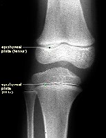
Long bones grow in length by interstitial growth of the epiphyseal plate and increase in thickness by appositional growth.
Lengthening of Long Bones
Epiphyseal Plate Zones
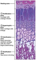
Interstitial growth requires epiphyseal cartilage. The epiphyseal plate consists of five zones:
Resting (quiescent) zone: Inactive cartilage.
Proliferation (growth) zone: Rapidly dividing cartilage.
Hypertrophic zone: Enlarged lacunae forming spaces.
Calcification zone: Matrix calcifies, chondrocytes die.
Ossification (osteogenic) zone: Spicules eroded and covered with new bone.
Epiphyseal Plate Closure
End of Bone Lengthening
Near the end of adolescence, the epiphyseal plate thins and is replaced by bone, ceasing bone lengthening. Epiphysis and diaphysis fuse, typically by age 18 in females and 21 in males.
Hormonal Regulation of Bone Growth
Key Hormones
Bone growth is regulated by:
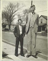
Growth Hormone: Stimulates epiphyseal plate activity.
Thyroid Hormone: Modulates growth hormone activity.
Testosterone & Estrogens: Promote growth spurt and induce epiphyseal plate closure.
Bone Remodeling
Resorption and Deposition
Bone remodeling involves resorption by osteoclasts and deposition by osteoblasts. Resorption breaks down bone matrix, while deposition lays down new osteoid matrix. Mechanical stress and calcium/phosphate concentrations may trigger remodeling.
Mechanical Stress on Bones
Bone Adaptation
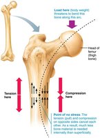
Bones adapt to mechanical stress, bending to compress one side and stretch the other. The diaphysis is thickest where most bending occurs, and bones can be hollow where compression and tension balance.
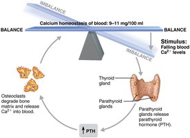
Calcium Homeostasis
Hormonal Regulation
Blood calcium levels are regulated by negative feedback loops:
Parathyroid Hormone (PTH): Stimulates bone resorption when calcium is low.
Calcitonin: Lowers blood calcium when levels are high (effects are negligible).
Bone Disorders
Common Pathologies
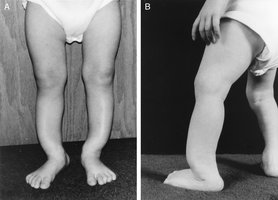
Osteomalacia: Poorly mineralized, soft bones due to lack of calcium/vitamin D.
Rickets: Childhood osteomalacia; severe due to growing bones.
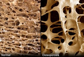
Osteoporosis: Resorption outpaces deposition, leading to brittle bones.
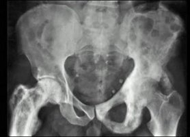
Paget’s Disease: Excessive, haphazard bone deposit and resorption; cause unknown.
Bone Fracture & Repair
Fracture Classification
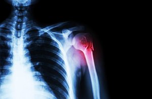
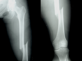
Fractures are classified by position of bone ends (nondisplaced/displaced), completeness (complete/incomplete), and skin penetration (open/closed).
Fracture Treatment and Repair
Treatment involves reduction (realignment) and immobilization. Repair occurs in four stages:
Hematoma formation
Fibrocartilaginous callus formation
Bony callus formation
Bone remodeling
Fracture Repair
Old fractures can be seen on x-rays due to ridges visible after repair.
Bone Disorder | Cause | Effect |
|---|---|---|
Osteomalacia | Lack of calcium/vitamin D | Soft, weak bones |
Rickets | Lack of calcium/vitamin D (children) | Severe bone deformity |
Osteoporosis | Resorption > deposition | Brittle, fracture-prone bones |
Paget's Disease | Unknown (possibly viral) | Deformed, painful bones |
Additional info: Academic context was added to clarify bone cell types, ossification processes, and hormonal regulation. All images included are directly relevant to the adjacent content and reinforce anatomical or physiological concepts.
