 Back
BackMuscle Contraction and Adaptation: Structure, Function, and Homeostasis
Study Guide - Smart Notes
 Tailored notes based on your materials, expanded with key definitions, examples, and context.
Muscle Contraction: Principles and Types
Whole Muscle Contraction
Muscle contraction is governed by the same principles in both single muscle fibers and whole muscles. The process produces muscle tension, which is the force exerted on a load or object to be moved. Contraction may or may not result in muscle shortening, depending on the type:
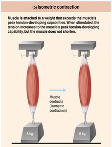
Isometric contraction: Muscle tension increases but does not exceed the load, so the muscle does not shorten.
Isotonic contraction: Muscle shortens because muscle tension exceeds the load.
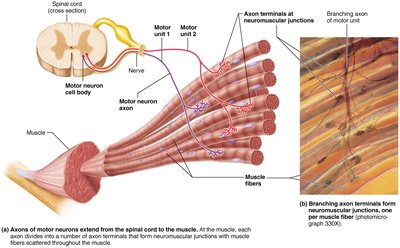
The Motor Unit
A motor unit consists of one motor neuron and all the muscle fibers it innervates. The number of fibers per motor unit can range from four to several hundred, with smaller numbers allowing greater fine control. Motor units are the functional nerve-muscle units, and their stimulation causes only weak contraction of the entire muscle due to the distribution of fibers throughout the muscle.
Each muscle is served by at least one motor nerve, which contains axons of many motor neurons.
Axons branch into terminals, forming neuromuscular junctions (NMJ) with individual muscle fibers.
Muscle Twitch and Graded Responses
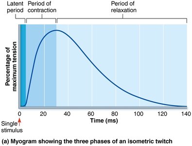
The Muscle Twitch
A muscle twitch is the simplest contraction, resulting from a muscle fiber’s response to a single action potential from a motor neuron. The twitch consists of three phases:
Latent period: Events of excitation-contraction coupling; no muscle tension is seen.
Period of contraction: Cross bridge formation; tension increases.
Period of relaxation: Ca2+ reentry into the sarcoplasmic reticulum (SR); tension declines to zero.
Muscle contracts faster than it relaxes. Differences in twitch strength and duration are due to variations in metabolic properties and enzymes between muscles.
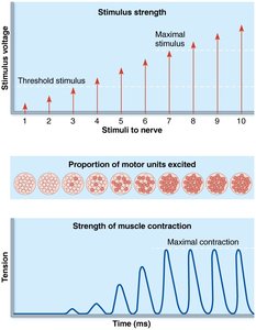
Graded Muscle Responses
Normal muscle contraction is smooth, and strength varies with needs. Graded muscle responses are required for proper control of skeletal movement and are achieved by:
Changing the frequency of stimulation
Changing the strength of stimulation
Muscle Tone
Muscle tone is the constant, slightly contracted state of all muscles, maintained by spinal reflexes. Groups of motor units are alternately activated in response to input from stretch receptors, keeping muscles firm, healthy, and ready to respond.
Types of Muscle Contractions
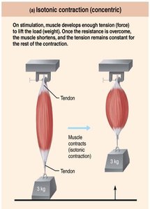
Isotonic and Isometric Contractions
Muscle contractions can be classified as isotonic or isometric:
Isotonic contractions: Muscle changes in length and moves a load. These can be concentric (muscle shortens and does work) or eccentric (muscle lengthens while generating force).
Isometric contractions: Muscle stays the same length; tension increases but the muscle does not shorten.
Examples:
Concentric: Biceps contract to pick up a book.
Eccentric: Laying a book down causes biceps to lengthen while generating force.
Isometric: Holding a book at arm’s length.
Energy for Muscle Contraction
ATP and Its Regeneration
ATP is the only source of energy for contractile activities and must be regenerated quickly. Muscle fibers use ATP to:
Move and detach cross bridges
Pump calcium back into the SR
Pump Na+ out and K+ back into the cell after excitation-contraction coupling
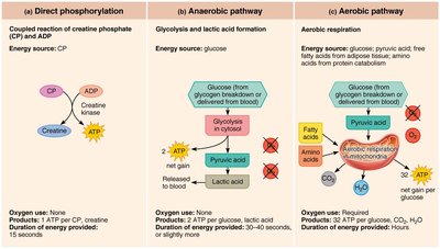
ATP is regenerated by three mechanisms:
Direct phosphorylation of ADP by creatine phosphate (CP)
Anaerobic pathway: glycolysis and lactic acid formation
Aerobic pathway
Comparison of Energy Sources During Exercise
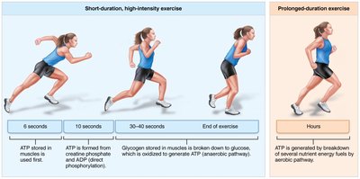
Different energy sources are used during short-duration and prolonged-duration exercise:
Short-duration, high-intensity exercise: ATP stored in muscles, creatine phosphate, and anaerobic glycolysis.
Prolonged-duration exercise: ATP generated by breakdown of several nutrient energy fuels by aerobic pathway.
Muscle Fatigue and Recovery
Muscle Fatigue
Muscle fatigue is the physiological inability to contract despite continued stimulation. Causes include:
Ionic imbalances (K+, Na+, Ca2+)
Increased inorganic phosphate (Pi) from CP and ATP breakdown
Decreased ATP and increased magnesium
Decreased glycogen
Lack of ATP is rarely a reason for fatigue, except in severely stressed muscles.
Excess Postexercise Oxygen Consumption (EPOC)
After exercise, muscles require extra oxygen to return to their pre-exercise state. This includes replenishing oxygen reserves, reconverting lactic acid to pyruvic acid, replacing glycogen stores, and resynthesizing ATP and creatine phosphate. This process is known as excess postexercise oxygen consumption (EPOC), formerly called "oxygen debt."
Velocity and Duration of Contraction
Factors Influencing Contraction
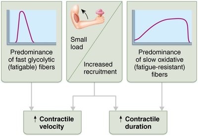
The speed and duration of muscle contraction are influenced by:
Muscle fiber type
Load
Recruitment
Muscle Fiber Types
Muscle fibers are classified based on:
Speed of contraction (slow or fast fibers)
Metabolic pathways used for ATP synthesis (oxidative or glycolytic)
Types of skeletal muscle fibers:
Slow oxidative fibers: Low-intensity, endurance activities (e.g., maintaining posture)
Fast oxidative fibers: Medium-intensity activities (e.g., sprinting or walking)
Fast glycolytic fibers: Short-term intense or powerful movements (e.g., hitting a baseball)
Most muscles contain a mixture of fiber types, resulting in a range of contractile speed and fatigue resistance. All fibers in one motor unit are the same type, and genetics dictate an individual’s percentage of each.
Adaptation to Exercise
Aerobic (Endurance) Exercise
Aerobic exercise, such as jogging, swimming, and biking, leads to increased muscle capillaries, number of mitochondria, and myoglobin synthesis. This results in greater endurance, strength, and resistance to fatigue, and may convert fast glycolytic fibers into fast oxidative fibers.
Resistance (Anaerobic) Exercise
Resistance exercise, such as weight-lifting or isometric exercises, leads to muscle hypertrophy (increase in fiber size), increased mitochondria, myofilaments, glycogen stores, and connective tissue, resulting in increased muscle strength and size.
Clinical and Homeostatic Imbalance
Disuse Atrophy
Muscles must be active to remain healthy. Disuse atrophy is the degeneration and loss of muscle mass due to immobilization or loss of neural stimulation. Muscle strength can decline 5% per day, and paralyzed muscles may atrophy to one-fourth their initial size. Fibrous connective tissue replaces lost muscle tissue, and rehabilitation becomes impossible at this stage.
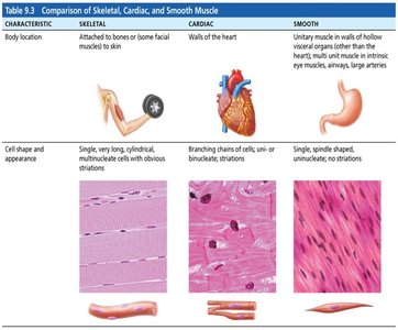
Comparison of Skeletal, Cardiac, and Smooth Muscle
Muscle Types
Skeletal, cardiac, and smooth muscles differ in location, cell shape, and appearance. The following table summarizes their characteristics:
Characteristic | Skeletal | Cardiac | Smooth |
|---|---|---|---|
Body location | Attached to bones or skin | Walls of the heart | Walls of hollow organs (except heart), blood vessels |
Cell shape and appearance | Long, cylindrical, multinucleate, striated | Branching chains, uni- or binucleate, striated | Single, spindle-shaped, uninucleate, no striations |
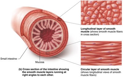
Smooth Muscle Contraction
Structure and Function
Smooth muscle is found in the walls of hollow organs and blood vessels. It consists of spindle-shaped cells with a single nucleus and no striations. Smooth muscle fibers are connected by gap junctions, allowing coordinated contraction. Dense bodies anchor intermediate filaments, and contraction occurs by sliding of actin and myosin filaments.
Developmental Aspects of Muscle
Muscle Repair and Aging
Skeletal muscle repair involves satellite cells and myoblast-like cells that help repair injured fibers and allow limited regeneration. Cardiac muscle is mostly repaired by scar tissue, while smooth muscle has good regenerative capacity. At birth, movements are uncoordinated and largely reflexive, developing neuromuscular coordination over time. With age, connective tissue increases, muscle fibers decrease, and muscle becomes more sinewy. By age 30, gradual loss of muscle mass (sarcopenia) begins.
References: Marieb, E. N., & Hoehn, K. (2023). Human anatomy and physiology (12th ed.). Pearson.
