 Back
BackMuscle Structure, Function, Physiology, and Pathology: Study Notes for Anatomy & Physiology
Study Guide - Smart Notes
 Tailored notes based on your materials, expanded with key definitions, examples, and context.
Muscle Structure and Naming
Muscle Naming Based on Shape
Muscles are often named according to their shape, which helps in identifying their anatomical location and function.
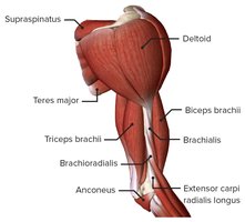
Deltoid: Named for its triangular shape.
Trapezius: Named for its trapezoid shape.
Rhomboid: Named for its rhombus shape.
Latissimus: Means 'wide'.
Teres: Means 'round'.
Rotator Cuff Muscles
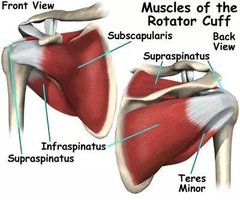
The rotator cuff is a group of muscles and their tendons that stabilize the shoulder.
Supraspinatus
Infraspinatus
Teres minor
Subscapularis
Quadriceps Muscle Group
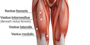
The quadriceps are the most powerful muscle group in the body, located in the anterior thigh.
Rectus Femoris
Vastus Medialis
Vastus Intermedius
Vastus Lateralis
Sternocleidomastoid Muscle (SCM)
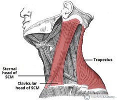
The SCM flexes and laterally rotates the head. It is located deep to the platysma on the anterolateral surface of the neck.
Action: Flexion and lateral rotation of the head.
Location: Anterolateral neck, deep to platysma.
Muscle Physiology and Response to Exercise
Muscle Adaptation to Exercise
Muscles adapt to regular exercise by increasing their size, number of mitochondria, and efficiency of oxygen utilization. This is known as the Overload Principle.
Hypertrophy: Increase in muscle fiber size and number.
Increased mitochondria: Enhanced energy production.
Improved O2 utilization: Greater endurance.
Muscle Tissue Resilience
Muscle tissue is highly vascular, providing nutrients and oxygen, and making it resistant to infection.
Rich blood supply: Facilitates nutrient delivery and waste removal.
Resistance to infection: Due to vascularity.
Hypertrophy Explained
Hypertrophy is the increase in size and number of muscle fibers, resulting from regular, increasing workloads. Damaged cells fuse, creating more contractile proteins and mitochondria.
Stronger, more efficient muscle tissue after recovery.
Muscle Shaking During Exercise
Muscle shaking occurs when motor units drop out due to fatigue, causing jerky contractions.
Motor units: Groups of muscle cells controlled by a neuron.
Asynchronous stimulation: Smooth function under normal conditions.
Fatigue: Leads to trembling contractions.
Muscle Soreness
Soreness is caused by micro-damage to muscle and connective tissue, leading to inflammation and pain. Lactic acid buildup may contribute.
Muscle Strain Injuries
Muscle strains are serious injuries caused by overstretching or involuntary contraction, resulting in tears and inflammation.
Risk factors: Dehydration, restricted blood supply, poor muscle tone, trauma.
Symptoms: Burning pain, nerve impingement.
Muscle Knots
Muscle knots are sensitive areas that remain contracted at rest, causing aching and hypersensitivity.
Causes: Over/underuse, poor posture, stress, diet.
Treatment: Stretching, heat/cold, massage, physical therapy.
Muscle Cramps
Muscle cramps are involuntary, forceful contractions that do not relax quickly.
Risk factors: Dehydration, low B vitamins, electrolyte imbalances, poor muscle tone, overuse, poor posture, nerve stimulation, circulatory problems, medications.
Anabolic Steroids and Muscle Growth
Anabolic steroids are variants of testosterone used to treat muscle wasting diseases and increase muscle mass, strength, and oxygen capacity.
Side effects: Acne, hair loss, infertility, liver damage, elevated cholesterol, psychiatric effects.
Sarcopenia and Muscle Atrophy
Sarcopenia is age-related muscle decline, where muscle fibers decrease and are replaced by connective tissue. Inactivity leads to muscle weakness and atrophy.
By age 80: Strength drops by 50%.
Disuse atrophy: Loss of muscle mass and strength due to immobilization.
Importance of Physical Activity
Staying active with resistance training throughout life is essential to maintain muscle mass and reduce fall risk.
Muscle Contraction Physiology
Temporal Summation and Recruitment
Muscle contraction strength is graded by frequency and strength of stimulation.
Temporal summation: Increased frequency of action potentials leads to stronger contraction.
Recruitment: Activation of more motor units increases contraction strength.
Muscle Tone
Relaxed muscles are always slightly contracted, known as muscle tone. This keeps muscles firm, healthy, and ready to respond.
Types of Muscle Contraction
Muscle contractions are categorized as isotonic or isometric.
Isotonic: Muscle changes length to move a load. Includes concentric (shortening) and eccentric (lengthening) contractions.
Isometric: Muscle tension develops, but the load is not moved.
Energy Pathways for Muscle Contraction
Muscle tissue requires ATP for contraction and other cellular functions. ATP is regenerated by three pathways:
Direct phosphorylation: Uses creatine phosphate for short bursts (15 seconds).
Anaerobic pathway: Breaks down glucose without oxygen, produces lactic acid (40 seconds).
Aerobic pathway: Uses oxygen, breaks down glucose to water and CO2, generates lots of ATP for prolonged activity.
Aerobic Endurance
Aerobic endurance is the length of time a muscle can contract using aerobic pathways. When demands exceed aerobic capacity, muscles switch to anaerobic pathways.
Factors Affecting Muscle Force
Muscle force depends on:
Frequency of stimulation
Number of fibers recruited
Size of fibers
Degree of muscle stretch
Degree of Muscle Stretch
Optimal sarcomere length allows maximal cross-bridge formation and force generation. Overstretched or contracted sarcomeres reduce force.
Velocity and Duration of Contraction
Muscles vary in contraction speed and endurance based on fiber type, load, and motor unit recruitment.
Slow twitch fibers: Endurance, slow fatigue, dominant in shoulders, forearms, lower legs.
Fast twitch fibers: Quick, powerful movements, fatigue quickly, dominant in chest, triceps, thighs, hamstrings.
Muscle Fatigue
Muscle fatigue occurs when tissue can no longer contract despite stimulation. Causes include ionic imbalances, increased phosphate, decreased ATP, increased Mg2+, and decreased glycogen.
Post-Exercise Recovery
After exercise, the body requires extra oxygen (EPOC) to restore ATP, glycogen, and myoglobin levels, repair proteins, and return temperature to normal.
Smooth Muscle
Structure and Function
Smooth muscle contracts slowly and synchronously, with sheets responding in unison. It is energy efficient and can maintain contraction for long periods.
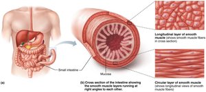
Innervation and Contraction
Innervated by autonomic nerve fibers, smooth muscle uses diffuse junctions and neurotransmitters (norepinephrine, ACh) to stimulate many cells.
Similarities to Skeletal Muscle
Uses Ca2+ influx for contraction
Actin and myosin cross-bridge formation
ATP required
Special Features
Spontaneous contraction when stretched
Stress-relaxation response allows hollow organs to fill without contraction
Muscle Pathology
Muscular Dystrophy
Muscular dystrophy is a group of inherited diseases causing muscle weakness and wasting. Types differ in affected muscles, age of onset, and progression.
Duchenne & Becker: Similar symptoms, Becker progresses slower.
Clinical presentation: Weakness, wasting, cardiac arrhythmias, cognitive impairments.
Diagnosis: Clinical suspicion, muscle biopsy.
Treatment: Supportive, steroids, physical therapy, cardiac monitoring.
Myasthenia Gravis
Myasthenia gravis is an autoimmune neuromuscular disease causing weakness and rapid fatigue of voluntary muscles, improving with rest.
Symptoms: Ptosis, difficulty speaking/swallowing, jaw fatigue, unbalanced facial expressions.
Pathophysiology: Antibodies block ACh receptors at NMJ.
Diagnosis: Blood tests, EMG.
Treatment: ACh-ase inhibitors, steroids, immunosuppressants.
Claudication
Claudication is pain in the legs during exercise due to insufficient arterial blood flow, improving with rest.
Risk factors: Smoking, diabetes, obesity, high blood pressure.
Diagnosis: Ultrasound.
Treatment: Medications, exercise, anti-platelet therapy.
Nemaline Myopathy
Nemaline myopathy is a rare, inherited disorder causing muscle weakness, poor tone, and diminished reflexes. It impairs cross-bridge formation.
Symptoms: Weakness, impaired swallowing, breathing, speech.
Treatment: Supportive care.
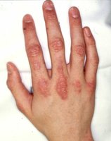
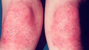
Polymyositis/Dermatomyositis
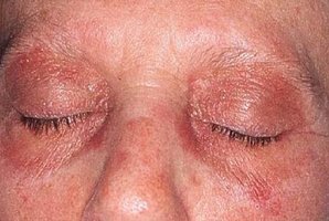
These are inflammatory muscle diseases, possibly viral or autoimmune, causing pain, stiffness, and progressive weakness.
Symptoms: Weakness in abdomen, shoulders, upper arms, hips; difficulty breathing/swallowing.
Diagnosis: Muscle biopsy.
Treatment: Steroids, supportive care.
Rhabdomyolysis
Rhabdomyolysis is a serious condition caused by extensive muscle damage, releasing myoglobin and other chemicals into the blood, damaging kidneys.
Causes: Overexertion, trauma, toxins, disease.
Symptoms: Weak, tender, painful muscles, urine changes.
Treatment: Fluid resuscitation, dialysis, monitoring for DIC.
Sarin Nerve Gas
Sarin is a toxic nerve agent that inhibits ACh-ase, causing continuous muscle contraction and fatal asphyxiation.
Botulism Infection
Botulism is caused by Clostridium botulinum toxin, which blocks ACh release, causing muscle weakness and paralysis.
Medical uses: Treatment of muscle spasms, migraines, hyperhidrosis, cosmetic procedures.
Risks: Eyelid droop, dizziness, facial weakness, difficulty swallowing, nausea.
Summary Table: Muscle Contraction Types
Type | Description | Example |
|---|---|---|
Isotonic (Concentric) | Muscle shortens to move a load | Picking up a book |
Isotonic (Eccentric) | Muscle lengthens while generating force | Thigh muscles during squats |
Isometric | Muscle tension develops, load not moved | Attempting to lift a piano |
Summary Table: Energy Pathways for Muscle Contraction
Pathway | Duration | Oxygen Required? | By-products |
|---|---|---|---|
Direct Phosphorylation | ~15 seconds | No | Creatine |
Anaerobic Glycolysis | ~40 seconds | No | Lactic acid |
Aerobic Respiration | Hours | Yes | CO2, H2O |
Summary Table: Muscle Fiber Types
Type | Contraction Speed | Fatigue Resistance | Primary Function |
|---|---|---|---|
Slow Twitch | Slow | High | Endurance |
Fast Twitch | Fast | Low | Powerful movements |
