 Back
Muscular System: Structure, Function, and Histology
Study Guide - Smart Notes
 Tailored notes based on your materials, expanded with key definitions, examples, and context.
Muscle Tissue Types
Overview of Muscle Tissue
Muscle tissue is specialized for contraction and is essential for movement, posture, and various bodily functions. There are three primary types of muscle tissue: skeletal, cardiac, and smooth muscle. Each type has distinct structural and functional characteristics.
Skeletal Muscle: Voluntary, striated, multinucleate cells attached to bones for movement.
Cardiac Muscle: Involuntary, striated, branched cells found in the heart, responsible for pumping blood.
Smooth Muscle: Involuntary, non-striated, spindle-shaped cells found in walls of hollow organs.
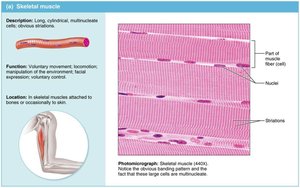
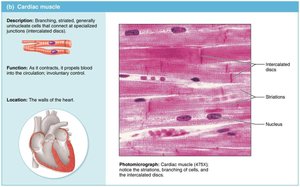
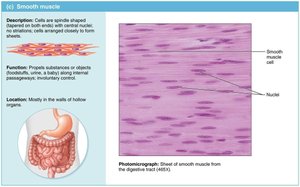
Comparison of Muscle Tissue Types
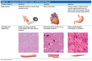
The three muscle types differ in location, cell shape, and function.
Characteristic | Skeletal | Cardiac | Smooth |
|---|---|---|---|
Body Location | Attached to bones or skin | Walls of the heart | Walls of hollow organs |
Cell Shape & Appearance | Long, cylindrical, multinucleate, striated | Branched, uninucleate, striated, intercalated discs | Spindle-shaped, uninucleate, no striations |
Control | Voluntary | Involuntary | Involuntary |
Skeletal Muscle Structure
Organization of Skeletal Muscle
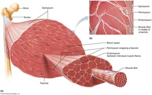
Skeletal muscle is organized into bundles of fibers, each surrounded by connective tissue layers.
Epimysium: Surrounds the entire muscle.
Perimysium: Surrounds each fascicle (bundle of muscle fibers).
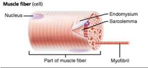
Endomysium: Surrounds individual muscle fibers.
Muscle Fiber: The basic contractile unit, containing myofibrils.
Myofibril and Sarcomere Structure
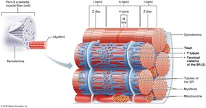
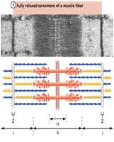
Myofibrils are composed of repeating units called sarcomeres, which are the functional units of muscle contraction.
Sarcomere: Defined by Z discs, contains thick (myosin) and thin (actin) filaments.
Striations: Result from the arrangement of actin and myosin filaments.
Key Regions: A band, I band, H zone, M line, Z disc.
Muscle Contraction Mechanisms
Excitation-Contraction Coupling
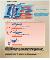
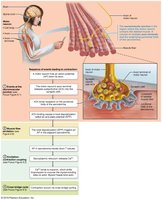
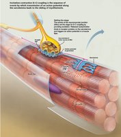
Excitation-contraction coupling is the process by which an action potential triggers muscle contraction.
Action Potential: Travels along the sarcolemma and into T-tubules.
Calcium Release: Ca2+ released from the sarcoplasmic reticulum binds to troponin, exposing binding sites on actin.
Cross-Bridge Formation: Myosin heads bind to actin, initiating contraction.
Sliding Filament Theory
Muscle contraction occurs as myosin heads pull actin filaments toward the center of the sarcomere, shortening the muscle.
ATP: Required for both contraction and relaxation.
Calcium: Regulates the interaction between actin and myosin.
Fascicle Arrangements in Muscles
Patterns of Fascicle Arrangement
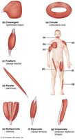
The arrangement of fascicles determines muscle shape and function.
Circular: Fascicles arranged in concentric rings (e.g., orbicularis oris).
Convergent: Broad origin, fascicles converge toward a single tendon (e.g., pectoralis major).
Parallel: Fascicles parallel to the long axis (e.g., sartorius).
Fusiform: Spindle-shaped with parallel fibers (e.g., biceps brachii).
Pennate: Short fascicles attach obliquely to a central tendon (e.g., rectus femoris). Types: unipennate, bipennate, multipennate.
Muscle Actions, Origins, and Insertions
Understanding Muscle Function
Muscles produce movement by contracting and pulling on bones. The origin is the fixed attachment, while the insertion moves with contraction.
Action: The movement produced by muscle contraction (e.g., flexion, extension).
Origin: The stationary attachment point.
Insertion: The movable attachment point.
Examples of Muscles and Their Actions
Latissimus Dorsi: Extension, adduction, and medial rotation of the arm.
Teres Major: Medial rotation and adduction of the arm.
Deltoid: Abduction of the arm.
Biceps Brachii: Flexion of the forearm at the elbow.
Gastrocnemius: Plantar flexion of the foot.
Summary Table: Major Muscles and Their Functions
Muscle | Action | Origin | Insertion |
|---|---|---|---|
Latissimus Dorsi | Extension, adduction, medial rotation | Spinous processes of lower vertebrae | Humerus |
Deltoid | Abduction | Clavicle, scapula | Deltoid tuberosity of humerus |
Biceps Brachii | Flexion of forearm | Scapula | Radius |
Gastrocnemius | Plantar flexion | Femur | Calcaneus |
Rectus Femoris | Extension of leg | Ilium | Patella, tibial tuberosity |
Soleus | Plantar flexion | Tibia, fibula | Calcaneus |
Sartorius | Flexion, abduction, lateral rotation | Ilium | Tibia |
Study Tips for Muscular System
Relate muscle names to their location and function.
Use anatomical models and diagrams to identify muscles.
Act out movements to understand muscle actions.
Practice labeling and identifying origins and insertions.
Key Terms and Definitions
Fascicle: Bundle of muscle fibers.
Myofibril: Rod-like unit within muscle fiber, composed of sarcomeres.
Sarcomere: Functional unit of muscle contraction.
Epimysium, Perimysium, Endomysium: Connective tissue layers surrounding muscle components.
Origin: Fixed attachment of muscle.
Insertion: Movable attachment of muscle.
Action: Movement produced by muscle contraction.
Additional info:
Muscle contraction is regulated by the nervous system, specifically motor neurons.
ATP is required for both contraction and relaxation of muscle fibers.
Calcium ions play a critical role in the regulation of contraction.
