 Back
BackNervous Tissue and Muscular System: Structure and Function
Study Guide - Smart Notes
 Tailored notes based on your materials, expanded with key definitions, examples, and context.
Nervous Tissue and Muscular System
Functions of the Nervous System
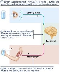
The nervous system is responsible for detecting changes in the environment, processing information, and initiating responses. It is essential for maintaining homeostasis and coordinating body activities.
Sensory Input: Sensory receptors detect stimuli from inside or outside the body and transmit this information to the central nervous system (CNS) via afferent pathways.
Integration: The CNS processes and interprets sensory input, deciding on an appropriate response.
Motor Output: The CNS sends signals via efferent pathways to effectors (muscles and glands) to produce a response.
Organization of the Nervous System
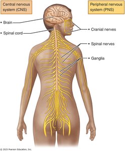
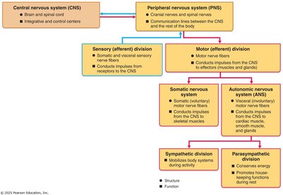
The nervous system is divided into two main parts: the central nervous system (CNS) and the peripheral nervous system (PNS).
Central Nervous System (CNS): Consists of the brain and spinal cord. It is the integration and control center, interpreting sensory input and dictating motor output.
Peripheral Nervous System (PNS): Includes all neural tissue outside the CNS, mainly nerves that extend from the brain and spinal cord (cranial and spinal nerves) and ganglia.
Divisions of the PNS
Sensory (Afferent) Division: Transmits impulses from sensory receptors to the CNS. Includes somatic sensory fibers (from skin, muscles, joints) and visceral sensory fibers (from organs).
Motor (Efferent) Division: Transmits impulses from the CNS to effectors. Subdivided into:
Somatic Nervous System: Voluntary control of skeletal muscles.
Autonomic Nervous System (ANS): Involuntary control of smooth muscle, cardiac muscle, and glands. Includes sympathetic (activates body systems) and parasympathetic (conserves energy) divisions.
Cell Types in Nervous Tissue
Neuroglia (Glial Cells)
Neuroglia are supporting cells that protect, insulate, and support neurons. Types include:
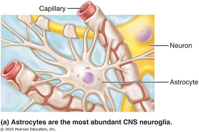
Astrocytes: Most abundant CNS glial cells; support neurons, regulate the chemical environment, and help form the blood-brain barrier.
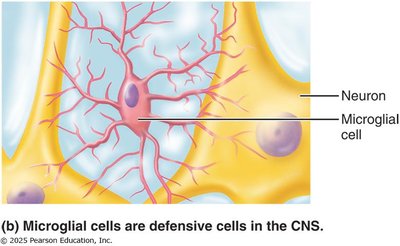
Microglial Cells: Act as phagocytes, removing debris and pathogens in the CNS.
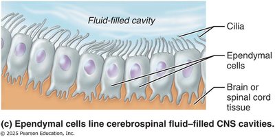
Ependymal Cells: Line cerebrospinal fluid-filled cavities; may be ciliated to help circulate CSF.
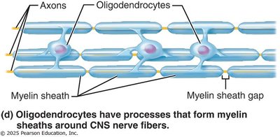
Oligodendrocytes: Form myelin sheaths around CNS nerve fibers.
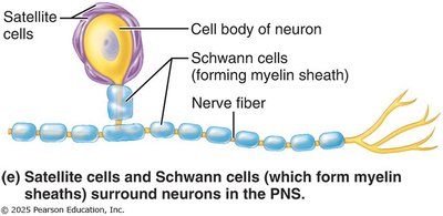
Satellite Cells (PNS): Surround neuron cell bodies in the PNS, similar to astrocytes.
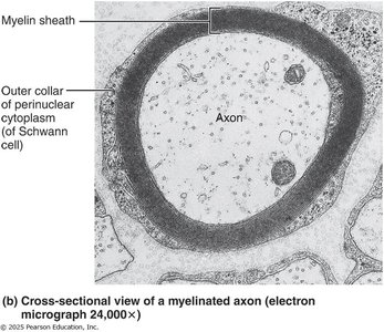
Schwann Cells (PNS): Form myelin sheaths around peripheral nerve fibers and assist in nerve regeneration.
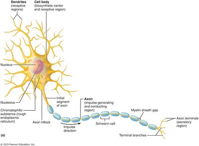
Neurons (Nerve Cells)
Neurons are the functional units of the nervous system, specialized for conducting electrical impulses. They have extreme longevity, are mostly amitotic, and have a high metabolic rate.
Cell Body (Soma): Contains the nucleus and organelles; biosynthetic and metabolic center.
Processes: Dendrites (receive input) and axons (transmit impulses).
Functional Classifications of Neurons
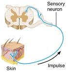
Sensory (Afferent) Neurons: Transmit impulses from receptors to the CNS; mostly unipolar; cell bodies in PNS ganglia.
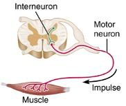
Motor (Efferent) Neurons: Carry impulses from CNS to effectors; multipolar; cell bodies in CNS.
Interneurons: Connect sensory and motor neurons within the CNS; most abundant type.
Structural Classifications of Neurons
Multipolar: Many processes (1 axon, many dendrites); most common in CNS.
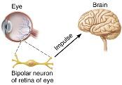
Bipolar: Two processes (1 axon, 1 dendrite); found in retina, ear, olfactory mucosa.
Unipolar (Pseudounipolar): One process that splits into two branches; mainly sensory neurons in PNS ganglia.
Myelin Sheath
The myelin sheath is a white, fatty covering that insulates axons, increasing the speed of nerve impulse transmission. Myelination differs between the CNS and PNS.
PNS Myelination: Formed by Schwann cells wrapping around axons. Gaps between Schwann cells are called nodes of Ranvier.
CNS Myelination: Formed by oligodendrocyte processes; one cell can myelinate multiple axons.
Function: Protects and insulates axons, speeds up electrical transmission.

Electrical Signals in Neurons
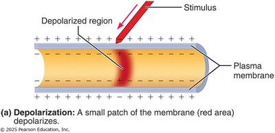
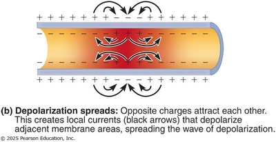
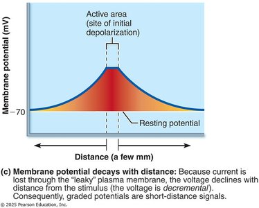
Graded Potentials
Graded potentials are short-lived, localized changes in membrane potential, essential for initiating action potentials. They occur in dendrites and cell bodies and decay with distance.
Types: Receptor potentials, postsynaptic potentials, end-plate potentials.
Mechanism: Triggered by stimulus opening gated ion channels; depolarization spreads but dissipates quickly.
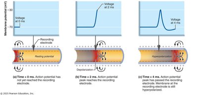
Action Potentials (APs)
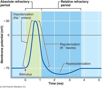
Action potentials are brief reversals of membrane potential that travel along axons, allowing long-distance communication. They are all-or-nothing events and do not decay with distance.
Phases: Depolarization (Na+ influx), repolarization (K+ efflux), hyperpolarization.
Threshold: Minimum depolarization needed to trigger an AP.
Propagation: APs are self-propagating and move in one direction along the axon.
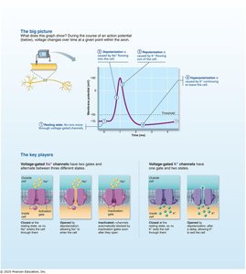
Refractory Periods
Absolute Refractory Period: No new AP can be generated; ensures one-way transmission.
Relative Refractory Period: AP can be generated only by a strong stimulus; some Na+ channels reset, K+ channels open.
Synapses and Neurotransmitters
Synapses
Synapses are junctions where neurons communicate with other neurons or effectors. They can be electrical (direct cytoplasmic connections) or chemical (neurotransmitter-mediated).
Presynaptic Neuron: Sends the signal.
Postsynaptic Neuron: Receives the signal.
Chemical Synapse Steps:
AP arrives at axon terminal.
Voltage-gated Ca2+ channels open.
Neurotransmitter released by exocytosis.
Neurotransmitter binds to postsynaptic receptors.
Ion channels open, creating graded potentials.
Neurotransmitter effects terminated.
Synaptic Delay: Time required for neurotransmitter release and binding; rate-limiting step.
Postsynaptic Potentials
Excitatory Postsynaptic Potentials (EPSP): Depolarize the postsynaptic membrane, increasing the likelihood of an AP.
Inhibitory Postsynaptic Potentials (IPSP): Hyperpolarize the membrane, decreasing the likelihood of an AP.
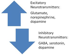
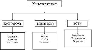
Neurotransmitters
Neurotransmitters are chemical messengers that transmit signals across synapses. They are classified by chemical structure and function.
Acetylcholine: First identified; used at neuromuscular junctions and by the ANS.
Biogenic Amines: Dopamine, norepinephrine, epinephrine, serotonin, histamine; involved in mood and behavior.
Amino Acids: Glutamate, aspartate, glycine, GABA.
Peptides: Substance P (pain), endorphins (natural opiates), gut-brain peptides.
Gases and Lipids: Nitric oxide, carbon monoxide, cannabinoids.
Muscular Tissue
Major Functions of Muscular Tissue
Muscular tissue is responsible for movement, posture, joint stabilization, and heat generation.
Produce Movement: Locomotion, manipulation, movement of substances.
Maintain Posture: Continuous muscle activity maintains posture.
Stabilize Joints: Muscles reinforce and stabilize joints.
Generate Heat: Muscle contractions produce heat, important for temperature regulation.
Skeletal Muscle Structure
Skeletal muscle is an organ composed of muscle fibers, connective tissue, blood vessels, and nerves. Each muscle fiber is covered by connective tissue sheaths: epimysium (entire muscle), perimysium (fascicles), and endomysium (individual fibers).
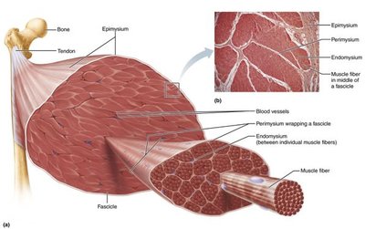
Skeletal Muscle Attachments
Direct Attachment: Epimysium fused to periosteum or perichondrium.
Indirect Attachment: Connective tissue extends as tendon or aponeurosis.
Skeletal Muscle Fibers
Muscle fibers are long, multinucleated cells with specialized structures:
Sarcolemma: Plasma membrane of muscle fiber.
Sarcoplasm: Cytoplasm containing glycosomes and myoglobin.
Myofibrils: Rod-like structures containing contractile proteins.
Sarcoplasmic Reticulum (SR): Stores and releases Ca2+.
T-Tubules: Invaginations of sarcolemma that transmit action potentials.
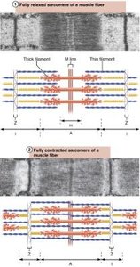
Myofilaments and Sarcomere Structure
Myofilaments are the contractile proteins of muscle fibers, organized into repeating units called sarcomeres.
Actin (Thin Filaments): Anchored to Z discs, extend across I band and part of A band.
Myosin (Thick Filaments): Extend the length of the A band, connected at the M line.
Other Proteins: Titin (elasticity), dystrophin (links to sarcolemma), regulatory proteins (tropomyosin, troponin).
Sarcomere: Smallest contractile unit, region between two Z discs.
Striations: Alternating dark (A bands) and light (I bands) regions.
Muscle Contraction Mechanism
Muscle contraction occurs through the sliding filament model, where myosin heads form cross-bridges with actin, pulling thin filaments toward the center of the sarcomere.
Cross Bridge Cycle: Attachment, power stroke, detachment, and cocking of myosin head (requires ATP).
Isotonic Contraction: Muscle changes length and moves a load (concentric or eccentric).
Isometric Contraction: Muscle tension increases but does not change length.
Muscle Twitch and Tetanus
Muscle Twitch: Response to a single action potential; consists of latent period, contraction, and relaxation phases.
Unfused (Incomplete) Tetanus: Increased stimulus frequency produces sustained, quivering contraction.
Fused (Complete) Tetanus: High-frequency stimulation produces smooth, sustained contraction; leads to fatigue.
Types of Muscle Tissue
Characteristic | Skeletal | Cardiac | Smooth |
|---|---|---|---|
Location | Attached to bones or skin | Walls of heart | Walls of hollow organs (except heart) |
Cell Shape | Long, cylindrical, multinucleate | Branching, striated, usually uninucleate | Spindle-shaped, uninucleate, non-striated |
Control | Voluntary | Involuntary | Involuntary |
Striations | Yes | Yes | No |
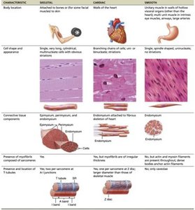
Smooth and Cardiac Muscle
Smooth Muscle: Found in walls of hollow organs; involuntary; organized in sheets; responsible for peristalsis and regulation of lumen diameter.
Cardiac Muscle: Found only in the heart; striated; involuntary; contracts at a steady rate set by pacemaker cells, modulated by the nervous system.
