 Back
BackNervous Tissue and Physiology: Structure and Function of the Nervous System
Study Guide - Smart Notes
 Tailored notes based on your materials, expanded with key definitions, examples, and context.
The Nervous System: Overview
Introduction to the Nervous System
The nervous system is a complex network of cells, tissues, and organs that enables communication within the body and with the external environment. It is responsible for sensory perception, integration of information, and motor responses necessary for survival and homeostasis.
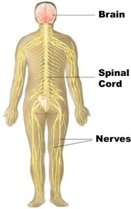
Organs of the Nervous System: Brain, spinal cord, nerves, and ganglia.
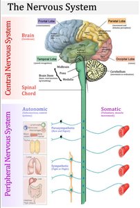
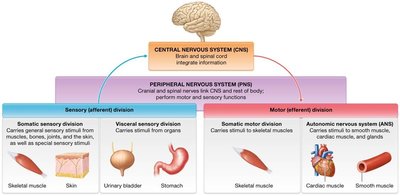
Major Divisions: Central Nervous System (CNS) and Peripheral Nervous System (PNS).
Anatomical Subdivisions of the Nervous System
Central Nervous System (CNS)
The CNS consists of the brain and spinal cord. It serves as the main control center, integrating sensory information and coordinating motor output.
Brain: Responsible for processing sensory information, initiating voluntary movements, and regulating autonomic functions.
Spinal Cord: Conducts signals between the brain and the rest of the body; involved in reflex actions.
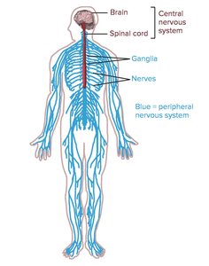
Peripheral Nervous System (PNS)
The PNS includes all neural tissue outside the CNS. It connects the CNS to limbs and organs, facilitating communication between the body and the brain.
Nerves: Bundles of axons that transmit signals to and from the CNS.
Ganglia: Clusters of neuron cell bodies located outside the CNS.
Functional Subdivisions of the Nervous System
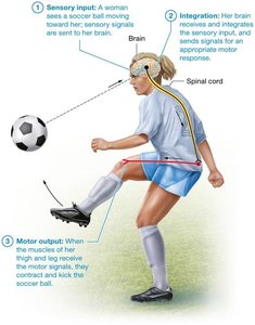
Three Primary Functions
The nervous system performs three essential functions: sensory input, integration, and motor output.
Sensory Input: Gathering information from sensory receptors about internal and external changes.
Integration: Processing and interpreting sensory input to determine an appropriate response.
Motor Output: Activating effector organs (muscles and glands) to produce a response.
Divisions of the Peripheral Nervous System
The PNS is divided into sensory (afferent) and motor (efferent) divisions, each with somatic and visceral components.
Sensory (Afferent) Division: Transmits sensory information to the CNS.
Somatic Sensory Fibers: Carry impulses from skin, skeletal muscles, and joints.
Visceral Sensory Fibers: Carry impulses from visceral organs.
Motor (Efferent) Division: Transmits commands from the CNS to effector organs.
Somatic Nervous System: Controls voluntary movements of skeletal muscles.
Autonomic Nervous System (ANS): Regulates involuntary functions (smooth muscle, cardiac muscle, glands).
Sympathetic Division: Prepares the body for 'fight or flight' responses.
Parasympathetic Division: Promotes 'rest and digest' activities.
Nervous Tissue Structure
Cellular Composition
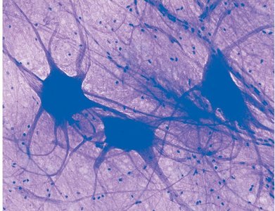
Nervous tissue is composed of two main cell types: neurons and neuroglia. Neurons are excitable cells responsible for transmitting electrical signals, while neuroglia support and protect neurons.
Neurons: Account for about 10% of nervous tissue cells but are the primary functional units.
Neuroglia: Make up about 90% of nervous tissue cells, providing structural and metabolic support.
Neuron Structure
Neurons have specialized structures that support their function in signal transmission.
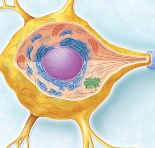
Cell Body (Soma): Contains the nucleus and organelles; responsible for metabolic activities.
Dendrites: Short, branched processes that receive signals from other neurons.
Axon: Long process that transmits action potentials away from the cell body.
Axon Hillock: Region where action potentials are initiated.
Myelin Sheath: Insulating layer that increases the speed of signal transmission.
Cell Body Details
The neuron cell body contains essential organelles for protein synthesis and energy production.
Nucleus: Contains genetic material.
Nissl Bodies: Clusters of rough endoplasmic reticulum and ribosomes for protein synthesis.
Mitochondria: Provide energy for cellular activities.
Neurofibrils: Intermediate filaments that maintain cell shape.
Myelination and Axonal Transport
Myelin Sheath Structure and Function
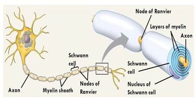
The myelin sheath is a multilayered lipid and protein covering that insulates axons, increasing the speed of electrical impulse conduction. Myelination is performed by different cell types in the CNS and PNS.
CNS: Oligodendrocytes form myelin sheaths around multiple axons.
PNS: Schwann cells form myelin sheaths around individual axons.
Nodes of Ranvier: Gaps in the myelin sheath that facilitate rapid signal transmission (saltatory conduction).
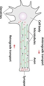
Axonal Transport
Axonal transport is the process by which materials are moved between the neuron cell body and the axon terminals. It is essential for neuron function and survival.
Anterograde Transport: Movement from the cell body toward the axon terminal (e.g., neurotransmitters, organelles).
Retrograde Transport: Movement from the axon terminal toward the cell body (e.g., recycled materials, signaling molecules).
Motor Proteins: Kinesin (anterograde) and dynein (retrograde) facilitate transport along microtubules.
Classification of Neurons
Structural Classification
Neurons are classified based on the number and arrangement of their processes.
Multipolar Neurons: One axon and multiple dendrites; most common type in the CNS.
Bipolar Neurons: One axon and one dendrite; found in sensory organs such as the retina.
Unipolar (Pseudounipolar) Neurons: Single process that splits into two branches; primarily sensory neurons in the PNS.

Functional Classification
Neurons are also classified by their function in the nervous system.
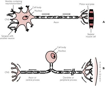
Sensory (Afferent) Neurons: Transmit impulses from sensory receptors to the CNS; mostly unipolar.
Motor (Efferent) Neurons: Carry impulses from the CNS to effectors (muscles/glands); mostly multipolar.
Interneurons: Connect sensory and motor neurons within the CNS; primarily multipolar.
Neuroglia: Supporting Cells
Types and Functions of Neuroglia
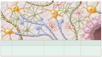
Neuroglia are non-neuronal cells that provide structural and functional support to neurons. There are six main types, four in the CNS and two in the PNS.
Neuroglia Type | Location | Function |
|---|---|---|
Astrocytes | CNS | Anchor neurons, maintain extracellular environment, form blood-brain barrier, repair tissue |
Oligodendrocytes | CNS | Form myelin sheaths around axons |
Microglia | CNS | Phagocytize debris and pathogens |
Ependymal Cells | CNS | Produce and circulate cerebrospinal fluid |
Schwann Cells | PNS | Form myelin sheaths around axons |
Satellite Cells | PNS | Support neuron cell bodies in ganglia |
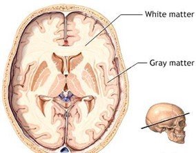
White Matter and Gray Matter
Composition and Appearance
The CNS contains regions of white matter and gray matter, distinguished by their composition and appearance.
White Matter: Composed mainly of myelinated axons; appears white due to the lipid content of myelin.
Gray Matter: Contains neuron cell bodies, unmyelinated dendrites, and axons; appears gray.
Myelin Sheath Formation and Axon Repair
Myelination in the CNS and PNS
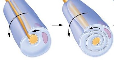
Myelination is the process by which glial cells wrap axons with myelin. The process differs between the CNS and PNS.
CNS: Oligodendrocytes wrap around multiple axons; no neurolemma is formed.
PNS: Schwann cells wrap around a single axon; the outermost layer forms the neurolemma, containing the Schwann cell nucleus and cytoplasm.
Axon Repair
Axon repair is possible in the PNS if the cell body and part of the myelinated axon remain intact. Schwann cells play a crucial role in guiding axonal regrowth. In the CNS, axon regeneration is limited due to the formation of scar tissue by astrocytes.
Wallerian Degeneration: The distal portion of the severed axon degenerates and is removed by phagocytes.
Regeneration Tube: Schwann cells and basal lamina form a tube to guide regrowth of the axon.
Growth Factors: Released by Schwann cells to stimulate axon regrowth.
Additional info: Neurons are generally amitotic (do not divide) and have a high metabolic rate, requiring continuous oxygen and glucose supply.
