 Back
Nervous Tissue: Structure, Function, and Neurophysiology
Study Guide - Smart Notes
 Tailored notes based on your materials, expanded with key definitions, examples, and context.
Nervous Tissue: Structure, Function, and Neurophysiology
Cellular Organization of the Nervous System
The nervous system is a complex network responsible for processing and transmitting information throughout the body. It is divided into three main anatomical and functional divisions: the Central Nervous System (CNS), Peripheral Nervous System (PNS), and Enteric Nervous System (ENS).
Central Nervous System (CNS): Composed of the brain and spinal cord; integrates, processes, and coordinates sensory and motor commands.
Peripheral Nervous System (PNS): Includes all nervous tissue outside the CNS, excluding the ENS.
Enteric Nervous System (ENS): Neural tissues in the wall of the gastrointestinal tract; controls digestive function.
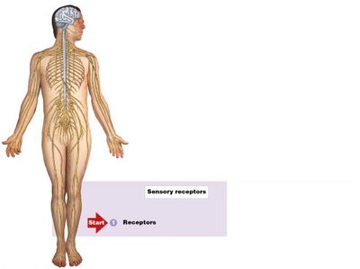
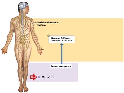
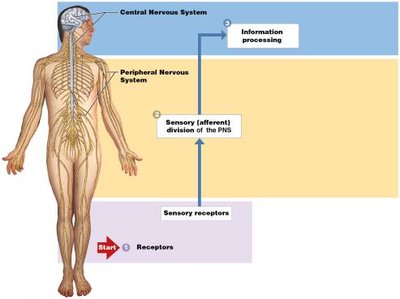
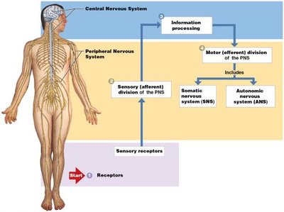
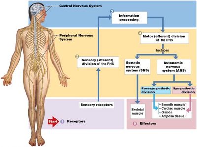
Functional Divisions of the PNS
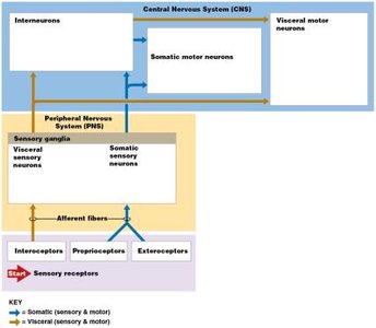
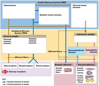
The PNS is further subdivided into sensory (afferent) and motor (efferent) divisions:
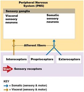
Sensory (afferent) division: Brings information to the CNS from receptors in peripheral tissues and organs.
Motor (efferent) division: Carries motor commands from the CNS to effectors. Includes the Somatic Nervous System (SNS) for voluntary control and the Autonomic Nervous System (ANS) for involuntary regulation.
General Functions of the Nervous System
The nervous system operates through a sequence of steps:
Receptors detect changes in the internal or external environment.
Information is sent to the CNS by the sensory division of the PNS.
Information processing occurs in the CNS.
Motor commands are carried by the motor division of the PNS.
Effectors respond to commands and change their activities.
Structure of a Typical Neuron
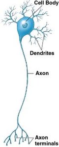
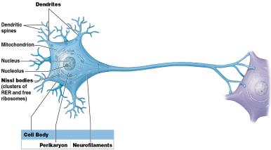
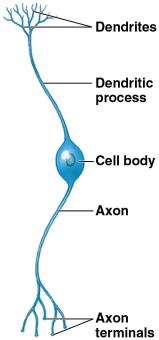
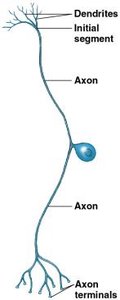
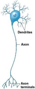
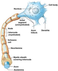
Neurons are specialized cells for intercellular communication. They have three main regions:
Dendrites: Receive stimuli from the environment or other neurons.
Cell Body (Soma): Contains the nucleus and organelles; site of metabolic activity.
Axon: Carries information toward other cells.
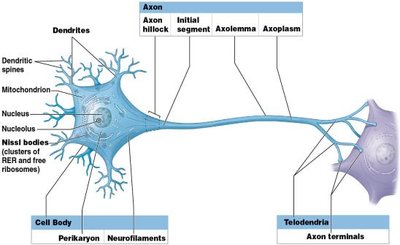
Axon Components and Synapses
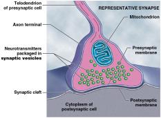
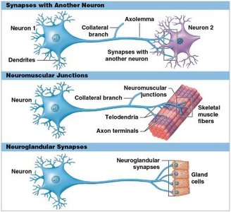
The axon includes the axon hillock (origin), initial segment (action potential initiation), axolemma (membrane), and axoplasm (cytoplasm). Axons end in telodendria and axon terminals, where synapses occur.
Synapse: The site where a neuron communicates with another cell, typically via neurotransmitter release into the synaptic cleft.
Neuron Classification by Structure
Neurons are classified anatomically into four types:
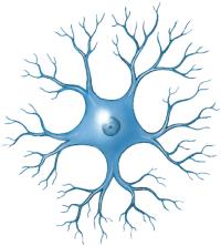
Anaxonic neurons: Small, indistinguishable axons and dendrites; found in brain and special sense organs.
Bipolar neurons: Two distinct processes; rare, found in special sense organs.
Unipolar neurons: Dendrites and axons are continuous; most sensory neurons in the PNS.
Multipolar neurons: Two or more dendrites and a single axon; most common in CNS and all motor neurons to skeletal muscles.
Neuron Classification by Function
Functionally, neurons are divided into:
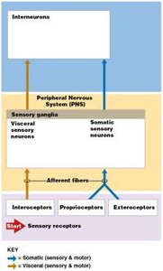
Sensory neurons: Detect stimuli and transmit information to the CNS.
Interneurons: Located in the CNS; connect sensory and motor neurons, responsible for higher functions.
Motor neurons: Carry commands from the CNS to effectors (muscles and glands).
Neuroglia of the CNS
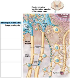
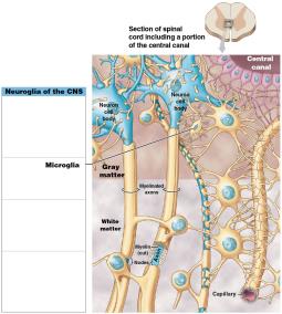
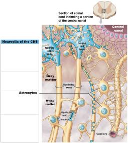
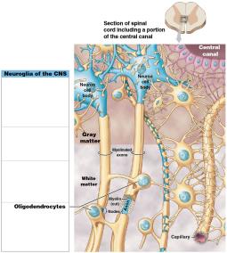
Neuroglia are supporting cells in the nervous system, comprising about half its volume. Four types are found in the CNS:
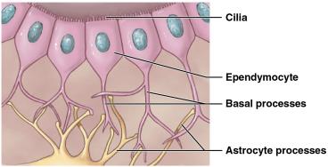
Ependymal cells: Line the central canal and ventricles; assist in producing and circulating cerebrospinal fluid (CSF).
Microglia: Mobile phagocytic cells; remove debris and pathogens.
Astrocytes: Maintain the blood-brain barrier, provide structural support, regulate ion/nutrient concentrations, absorb/recycle neurotransmitters, and form scar tissue after injury.
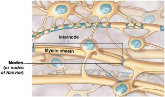
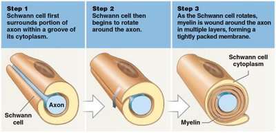
Oligodendrocytes: Stabilize axons and produce myelin sheaths, speeding up nerve impulse transmission.
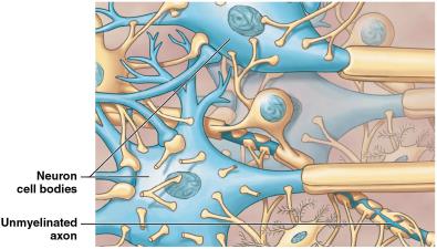
Neuroglia of the PNS
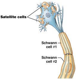
Two types of neuroglia are found in the PNS:
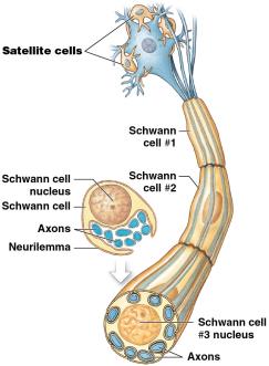
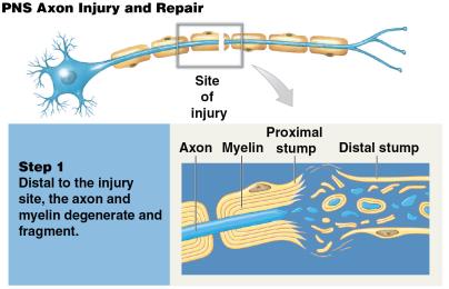
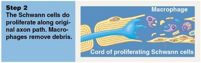
Schwann cells: Cover peripheral axons, participate in axon repair, and form myelin sheaths.
Satellite cells: Surround peripheral cell bodies and regulate the environment around neurons.
Neurophysiology: Membrane Potentials and Neuronal Activity
Neuronal activity depends on changes in membrane potential, which is the potential difference across a membrane due to unequal charge distribution.
Resting membrane potential: The membrane potential of an undisturbed cell, typically around –70 mV in neurons.
Graded potential: Temporary, localized change in resting potential produced by a stimulus.
Action potential: Electrical event that spreads along the axon surface toward axon terminals.
Synaptic activity: Neurotransmitter release changes permeability and produces graded potentials in the postsynaptic cell.
Creation and Maintenance of Resting Membrane Potential
The resting membrane potential is determined by differences in ion concentrations and permeability:
Extracellular fluid: High in Na+ and Cl–.
Cytosol: High in K+ and negatively charged proteins.
Leak channels: Allow passive diffusion of Na+ and K+.
Sodium–potassium exchange pump: Ejects 3 Na+ for every 2 K+ recovered, maintaining the potential.
Ion | Equilibrium Potential |
|---|---|
Potassium (K+) | –90 mV |
Sodium (Na+) | +66 mV |
Gated Ion Channels
Gated channels change membrane permeability in response to stimuli. Three types exist:
Chemically gated channels: Open when binding specific chemicals (e.g., acetylcholine).
Voltage-gated channels: Open/close in response to changes in membrane potential; characteristic of excitable membranes.
Mechanically gated channels: Open in response to physical distortion of the membrane.
Graded Potentials
Graded potentials are localized changes in membrane potential caused by stimuli that open gated ion channels. They decrease with distance from the stimulus and are proportional to stimulus size.
Depolarization: Shift toward a more positive membrane potential.
Repolarization: Return to resting membrane potential.
Hyperpolarization: Shift past resting membrane potential (more negative).
Action Potentials
Action potentials are all-or-none events for communication that begin with membrane potential reversal. They depend on voltage-gated ion channels and allow long-range communication.
Depolarization to threshold (about –60 mV).
Activation of sodium channels; rapid depolarization.
Inactivation of sodium channels; activation of potassium channels; repolarization starts.
Potassium channels close; brief hyperpolarization.
Refractory periods: Absolute (no response possible) and relative (response possible to stronger stimulus).
Propagation of Action Potentials
Action potentials propagate along axons by two mechanisms:
Continuous propagation: Step-by-step movement in unmyelinated axons; slower.
Saltatory propagation: Occurs in myelinated axons; depolarization only at nodes of Ranvier; faster.
Synapses and Information Transfer
Synapses are sites where information is transferred from one neuron to another or to an effector cell. Two types exist:
Chemical synapse: Most common; relies on neurotransmitter release (e.g., acetylcholine).
Electrical synapse: Rare; presynaptic and postsynaptic membranes joined by gap junctions.
Postsynaptic Potentials and Information Processing
Postsynaptic potentials are graded potentials in response to neurotransmitters:
Excitatory postsynaptic potential (EPSP): Graded depolarization; shifts membrane potential closer to threshold.
Inhibitory postsynaptic potential (IPSP): Graded hyperpolarization; shifts membrane potential farther from threshold.
Summation integrates effects of EPSPs and IPSPs:
Temporal summation: Repeated stimulation at a single synapse.
Spatial summation: Multiple synapses active simultaneously.
Higher Levels of Information Processing
Regulatory neurons and neurotransmitters facilitate or inhibit activities of presynaptic neurons. Over 100 neurotransmitters exist, each with direct or indirect effects on ion channels. Information is encoded as action potential frequency.
Key Equations:
Resting membrane potential:
Sodium-potassium pump:
Equilibrium potential (Nernst equation):
Additional info: The notes above expand on brief lecture points, providing definitions, examples, and context for each concept. Images included are directly relevant to the anatomical and physiological explanations of nervous tissue.
