 Back
BackNervous Tissue: Structure, Function, and Physiology
Study Guide - Smart Notes
 Tailored notes based on your materials, expanded with key definitions, examples, and context.
Nervous Tissue
Introduction to the Nervous System
The nervous system is responsible for receiving, processing, and responding to internal and external stimuli. It consists of the brain, spinal cord, sensory receptors, and nerves that connect to other body systems. The nervous system's main functions include:
Receiving information from sensory receptors
Processing and integrating sensory data
Initiating responses through motor commands
Nervous tissue contains two main cell types:
Neurons: Specialized for intercellular communication
Neuroglia (glial cells): Support, protect, and maintain the structure of nervous tissue
Divisions of the Nervous System
The nervous system is divided both anatomically and functionally:
Central Nervous System (CNS): Brain and spinal cord; integrates, processes, and coordinates sensory information and motor commands. Responsible for higher functions such as intelligence, memory, learning, and emotion.
Peripheral Nervous System (PNS): All nervous tissue outside the CNS; delivers sensory information to the CNS and carries motor commands to peripheral tissues. Includes cranial and spinal nerves.
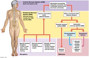
The PNS is further divided functionally:
Afferent division: Carries sensory information from receptors to the CNS.
Efferent division: Carries motor commands from the CNS to effectors (muscles, glands, adipose tissue).
The efferent division includes:
Somatic Nervous System (SNS): Controls voluntary and involuntary (reflex) skeletal muscle contractions.
Autonomic Nervous System (ANS): Controls involuntary actions of smooth muscle, cardiac muscle, glands, and adipose tissue. Subdivided into:
Sympathetic division: Prepares the body for 'fight or flight' responses.
Parasympathetic division: Promotes 'rest and digest' activities.
Enteric Nervous System (ENS): Neurons in the digestive tract that coordinate local reflexes independently of the CNS, but can be influenced by the ANS.
Neurons
Structure of a Typical Neuron
Neurons are the basic functional units of the nervous system, specialized for communication, information processing, and control. They have an excitable plasma membrane and can send and receive signals.
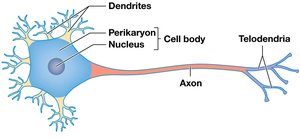
Dendrites: Receive incoming signals
Cell body (soma): Contains the nucleus and organelles
Axon: Conducts electrical impulses (action potentials) away from the cell body
Telodendria: Terminal branches that form synapses with other cells
Classification of Neurons
Neurons can be classified by structure and function:
Structural classification:
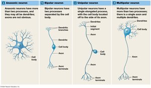
Anaxonic neurons: No obvious axon; found in the brain and special sense organs
Bipolar neurons: Two processes separated by the cell body; found in special sensory organs
Unipolar neurons: Single elongated process with the cell body off to the side; most sensory neurons of the PNS
Multipolar neurons: Many dendrites, one axon; most common type in the CNS
Functional classification:
Sensory (afferent) neurons: Carry information from receptors to the CNS
Motor (efferent) neurons: Carry instructions from the CNS to effectors
Interneurons: Integrate sensory information and coordinate motor output; found in the CNS
Sensory receptors are further classified as:
Interoceptors: Monitor internal systems and sensations
Exteroceptors: Monitor external environment (touch, temperature, etc.)
Proprioceptors: Monitor position and movement of muscles and joints
Neuroglia
Types and Functions of Neuroglia
Neuroglia are supporting cells that protect and maintain neurons. They make up about half the volume of the nervous system.
Central Nervous System (CNS) | Peripheral Nervous System (PNS) |
|---|---|
Astrocytes Ependymal cells Oligodendrocytes Microglia | Satellite cells Schwann cells |
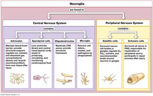
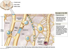
Astrocytes: Maintain blood-brain barrier, provide structural support, regulate ion and nutrient concentrations, absorb and recycle neurotransmitters, form scar tissue after injury
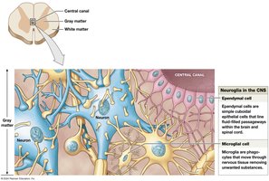
Ependymal cells: Line ventricles and central canal, produce and circulate cerebrospinal fluid
Oligodendrocytes: Myelinate CNS axons, provide structural framework
Microglia: Remove cell debris, wastes, and pathogens by phagocytosis
Satellite cells: Surround neuron cell bodies in ganglia, regulate environment
Schwann cells: Myelinate PNS axons, assist in repair after injury
Membrane Potential
Resting Membrane Potential
The resting membrane potential is the electrical potential difference across the plasma membrane of an unstimulated neuron, typically around -70 mV. All neural activity begins with a change in this potential.
Chemical gradient: Ions move passively across the membrane due to concentration differences
Electrical gradient: Ions move due to charge differences across the membrane
Electrochemical gradient: The sum of chemical and electrical forces acting on an ion
The sodium-potassium exchange pump (Na+/K+ ATPase) maintains the resting potential by moving 3 Na+ out and 2 K+ into the cell per ATP hydrolyzed.
Membrane Channels
Changes in membrane potential result from the opening or closing of specific ion channels:
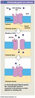
Chemically gated (ligand-gated) channels: Open in response to binding of a chemical messenger (e.g., neurotransmitter)
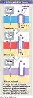
Voltage-gated channels: Open or close in response to changes in membrane potential
Mechanically gated channels: Open or close in response to physical distortion of the membrane
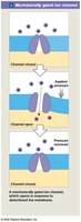
Graded Potentials
Graded potentials are temporary, localized changes in the membrane potential caused by the opening of gated ion channels. They can result in:
Depolarization: Membrane potential becomes less negative (e.g., Na+ influx)
Hyperpolarization: Membrane potential becomes more negative (e.g., K+ efflux)
Repolarization: Return to resting membrane potential after depolarization
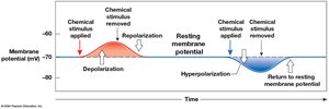
Action Potentials
Generation and Propagation of Action Potentials
An action potential is a large, rapid depolarization that propagates along the axon without diminishing. It follows the all-or-none principle: it either occurs fully or not at all.
Step 1: Depolarization to threshold – Graded potential depolarizes the initial segment to threshold
Step 2: Rapid depolarization – Voltage-gated Na+ channels open, Na+ enters
Step 3: Repolarization – At +30 mV, Na+ channels inactivate, K+ channels open, K+ exits
Step 4: Hyperpolarization – K+ channels close slowly, causing a brief hyperpolarization before returning to resting potential
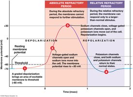
The refractory period is the time during which the neuron cannot (absolute) or can only with a strong stimulus (relative) generate another action potential.
Propagation of Action Potentials
Continuous propagation: Occurs in unmyelinated axons; action potential moves stepwise along the axon
Saltatory propagation: Occurs in myelinated axons; action potential jumps from node to node, increasing speed and efficiency
Propagation speed: Increased by myelination and larger axon diameter
Synapses
Structure and Function of Synapses
A synapse is a specialized site where a neuron communicates with another cell. The presynaptic cell sends the message, and the postsynaptic cell receives it.
Electrical synapses: Direct physical contact via gap junctions; rapid transmission
Chemical synapses: Use neurotransmitters to transmit signals across a synaptic cleft; most common type
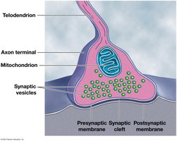
Types of chemical synapses include axoaxonic, axosomatic, axodendritic, neuromuscular, and neuroglandular junctions.
Events at a Cholinergic Synapse
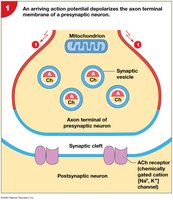
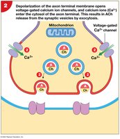
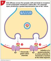
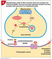
Cholinergic synapses release acetylcholine (ACh). The sequence of events is:
An action potential arrives at the axon terminal and depolarizes the membrane
Voltage-gated Ca2+ channels open, Ca2+ enters, triggering exocytosis of ACh
ACh binds to receptors on the postsynaptic membrane, causing Na+ influx and a graded potential
ACh is broken down by acetylcholinesterase (AChE), and byproducts are removed
Neurotransmitters and Neuromodulators
Classes and Effects
Neurotransmitters can be classified by their effects:
Excitatory: Cause depolarization and promote action potentials
Inhibitory: Cause hyperpolarization and suppress action potentials
The effect depends on the receptor, not the neurotransmitter itself.
Acetylcholine (ACh): Excitatory; found in CNS and PNS
Biogenic amines: Norepinephrine (NE), dopamine, serotonin
Amino acids: Glutamate (excitatory), glycine (inhibitory), GABA (inhibitory)
Neuropeptides: Opioids (enkephalins, endorphins, dynorphins)
Dissolved gases: Nitric oxide (NO), carbon monoxide (CO)
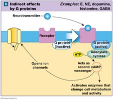
Neurotransmitters and neuromodulators act via:
Ionotropic effects: Directly open/close ion channels (e.g., ACh, glutamate)
Metabotropic effects: Indirectly affect the cell via G protein-coupled receptors and second messengers (e.g., NE, dopamine, serotonin, GABA)
Information Processing in Nervous Tissue
Integration and Summation
Postsynaptic cells integrate thousands of incoming signals. The net effect at the axon hillock determines whether an action potential is generated.
Excitatory postsynaptic potential (EPSP): Graded depolarization
Inhibitory postsynaptic potential (IPSP): Graded hyperpolarization
Summation: Integration of all incoming graded potentials
Temporal summation: Rapid, repeated stimuli at a single synapse
Spatial summation: Simultaneous stimuli at multiple synapses
The frequency of action potentials encodes the strength of the stimulus. Neuromodulators and other chemicals can facilitate or inhibit neuronal activity, altering the response of postsynaptic neurons.
