 Back
BackNeural Tissue: Structure and Function in the Nervous System
Study Guide - Smart Notes
 Tailored notes based on your materials, expanded with key definitions, examples, and context.
Introduction to the Nervous System
Overview and Functional Significance
The nervous system is one of the primary control and communication systems of the body, working alongside the endocrine system to regulate and coordinate bodily functions. It is characterized by rapid, brief responses (in contrast to the slower, longer-lasting effects of the endocrine system) and is responsible for processing sensory information, integrating data, and initiating motor responses.
Chemical communication is a shared feature with the endocrine system, but the nervous system uses electrical impulses for swift signaling.
The nervous system is divided into the central nervous system (CNS) and the peripheral nervous system (PNS).
Anatomical and Functional Divisions of the Nervous System
Central and Peripheral Nervous Systems
The nervous system is organized into two main anatomical divisions, each with distinct roles:
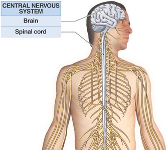
Central Nervous System (CNS): Consists of the brain and spinal cord. It is the main center for processing, integrating, and coordinating sensory data and motor commands. The CNS is also responsible for higher functions such as intelligence, memory, learning, and emotion.
Peripheral Nervous System (PNS): Composed of all neural tissue outside the CNS. It delivers sensory information to the CNS and carries motor commands to peripheral tissues and systems.
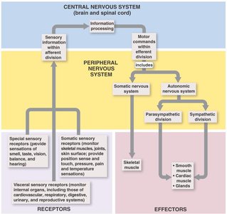
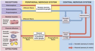
Functional Subdivisions of the PNS
Afferent Division: Brings sensory information from receptors to the CNS. Receptors can be somatic (skeletal muscles, joints, skin), visceral (internal organs), or special sense organs (eyes, ears, nose, tongue).
Efferent Division: Carries motor commands from the CNS to effectors (muscles and glands). It is further divided into:
Somatic Nervous System (SNS): Controls voluntary and involuntary skeletal muscle contractions.
Autonomic Nervous System (ANS): Regulates involuntary actions in smooth muscle, cardiac muscle, and glands. The ANS is subdivided into sympathetic and parasympathetic divisions.
Cellular Organization in Neural Tissue
Neurons and Neuroglia
Neural tissue is composed of two main cell types: neurons and neuroglia (glial cells).
Neurons: Specialized for the transfer and processing of information. They are excitable cells capable of generating and conducting electrical impulses.
Neuroglia: Supporting cells that provide structural framework, maintain the intercellular environment, and act as phagocytes. Neuroglia outnumber neurons by about five to one.
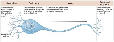
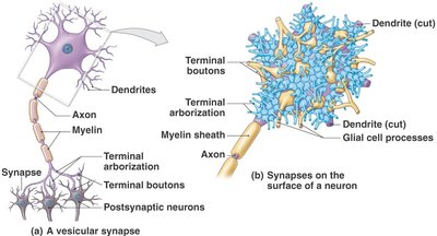
Structure of a Typical Neuron
Dendrites: Receive stimuli from the environment or other neurons.
Cell Body (Soma): Contains the nucleus and organelles; integrates incoming signals.
Axon: Conducts nerve impulses (action potentials) away from the cell body toward synaptic terminals.
Terminal Boutons: Affect other neurons or effector organs (muscles or glands).
Types of Neuroglia
Neuroglia are classified based on their location and function:
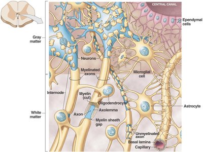
Central Nervous System (CNS): Astrocytes, oligodendrocytes, microglia, and ependymal cells.
Peripheral Nervous System (PNS): Satellite cells and Schwann cells.
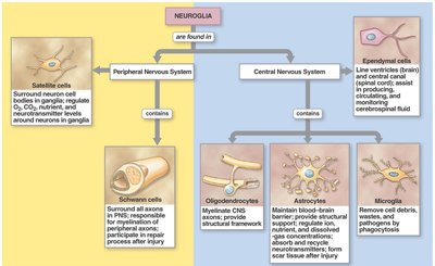
Functions of Major Neuroglial Cells
Astrocytes: Maintain the blood-brain barrier, provide structural support, regulate ion and nutrient concentrations, and repair damaged tissue.
Oligodendrocytes: Myelinate CNS axons, increasing the speed of impulse conduction.
Microglia: Act as phagocytes, removing debris and pathogens.
Ependymal Cells: Line ventricles and central canal, producing and circulating cerebrospinal fluid.
Satellite Cells (PNS): Surround neuron cell bodies in ganglia, regulating the environment.
Schwann Cells (PNS): Myelinate peripheral axons and assist in axon regeneration after injury.
Classification of Neurons
Structural and Functional Types
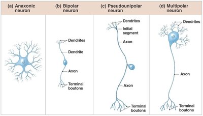
Structural Classification: Based on the number of processes extending from the cell body:
Anaxonic Neurons: No obvious axon; found in the brain and special sense organs.
Bipolar Neurons: One dendrite and one axon; found in sensory organs (e.g., retina).
Pseudounipolar Neurons: Single process that splits into two branches; most sensory neurons in the PNS.
Multipolar Neurons: Many dendrites, one axon; most common type, including motor neurons.
Functional Classification:
Sensory Neurons: Transmit sensory information from receptors to the CNS.
Motor Neurons: Carry commands from the CNS to effectors (muscles/glands).
Interneurons: Connect sensory and motor neurons within the CNS; involved in processing and integration.
Receptors and Effectors
Types of Receptors
Exteroceptors: Monitor external environment (touch, temperature, pressure, special senses).
Proprioceptors: Monitor position and movement of skeletal muscles and joints.
Interoceptors: Monitor internal environment (digestive, respiratory, cardiovascular, urinary, reproductive systems).
Neural Regeneration
Regeneration of Peripheral Nerves
Peripheral nerves have a limited ability to regenerate after injury, primarily due to the activity of Schwann cells. The process involves the degeneration of the distal axon, proliferation of Schwann cells, and regrowth of the axon along the original path.
Nerve Impulse and Synaptic Communication
Action Potentials and Conduction
Excitability is the ability of a neuron's plasma membrane to conduct electrical impulses. An action potential is an electrical impulse that travels along the axon. The speed of conduction depends on the presence of a myelin sheath and the diameter of the axon.
Myelinated axons conduct impulses faster than unmyelinated axons.
Larger diameter axons conduct impulses more rapidly.
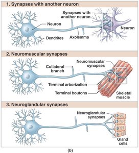
Synaptic Communication
Neurons communicate with other neurons, muscle cells, or gland cells at specialized junctions called synapses. Synapses can be electrical or chemical (vesicular), with the latter involving neurotransmitter release.
Synapses with Neurons: Transfer signals between neurons.
Neuromuscular Synapses: Transfer signals from neurons to muscle fibers.
Neuroglandular Synapses: Transfer signals from neurons to gland cells.
Organization of Neural Circuits
Types of Neural Circuits
Neural circuits are patterns of synaptic connections that determine the flow of information within the nervous system. Major types include:
Divergence: One neuron sends signals to multiple neurons.
Convergence: Multiple neurons send signals to a single neuron.
Serial Processing: Neurons are arranged in a linear sequence.
Parallel Processing: Information is processed simultaneously along multiple pathways.
Reverberation: Feedback loops maintain activity within the circuit.
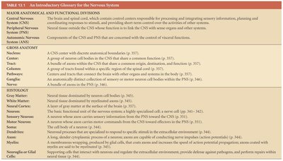
Glossary of Key Terms
Term | Definition |
|---|---|
Central Nervous System (CNS) | Brain and spinal cord; main processing and integration center. |
Peripheral Nervous System (PNS) | Neural tissue outside the CNS; connects CNS to limbs and organs. |
Neuron | Excitable cell that transmits electrical signals. |
Neuroglia | Supporting cells that protect, insulate, and nourish neurons. |
Gray Matter | Regions dominated by neuron cell bodies. |
White Matter | Regions dominated by myelinated axons. |
Ganglion | Collection of neuron cell bodies in the PNS. |
Nucleus (CNS) | Collection of neuron cell bodies in the CNS. |
Tract | Bundle of axons in the CNS. |
Nerve | Bundle of axons in the PNS. |
Summary
The nervous system is a complex network responsible for rapid communication and control throughout the body. Its organization into central and peripheral divisions, the diversity of neurons and neuroglia, and the mechanisms of signal transmission and processing are foundational concepts for understanding human anatomy and physiology.
