 Back
BackNeurons: Cellular and Network Properties
Study Guide - Smart Notes
 Tailored notes based on your materials, expanded with key definitions, examples, and context.
Neurons: Cellular and Network Properties
Introduction to the Nervous System
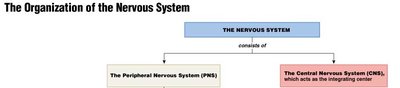
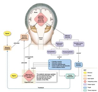
The nervous system is a complex network responsible for coordinating voluntary and involuntary actions and transmitting signals throughout the body. It is divided into the central nervous system (CNS) and the peripheral nervous system (PNS), each with specialized roles in processing and relaying information.
Organization of the Nervous System
Central Nervous System (CNS): Consists of the brain and spinal cord, serving as the main integrating and processing center.
Peripheral Nervous System (PNS): Composed of nerve tissue outside the CNS, including cranial nerves, spinal nerves, ganglia, plexuses, and sensory receptors.
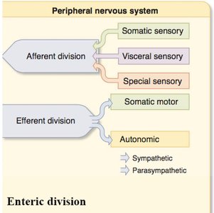
Divisions of the PNS
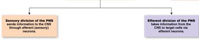
Afferent (Sensory) Division: Sends information from sensory receptors to the CNS.
Efferent (Motor) Division: Transmits commands from the CNS to effectors (muscles and glands).
Autonomic Division: Controls involuntary functions and is further divided into sympathetic and parasympathetic branches.
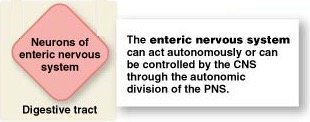
Enteric Division: A network of neurons in the digestive tract that can function independently or under CNS control.
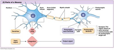
Neurons: Structure and Function
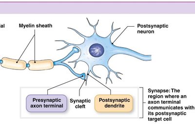
Neurons are the basic signaling units of the nervous system, specialized for the conduction of electrical impulses. They consist of a cell body (soma), dendrites, and an axon. Dendrites receive incoming signals, while axons transmit outgoing signals to target cells.
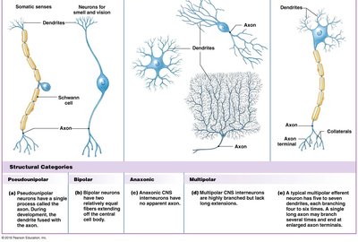
Structural Categories of Neurons
Pseudounipolar: Single process that splits into two branches; common in sensory neurons.
Bipolar: Two processes extending from the cell body; found in sensory organs.
Anaxonic: No apparent axon; found in the CNS.
Multipolar: Many dendrites and a single axon; typical of CNS interneurons and motor neurons.
Functional Categories of Neurons
Sensory (Afferent) Neurons: Transmit sensory information to the CNS.
Interneurons: Facilitate communication within the CNS.
Efferent Neurons: Carry signals from the CNS to effectors (muscles and glands).
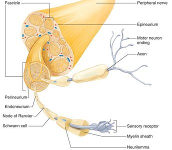
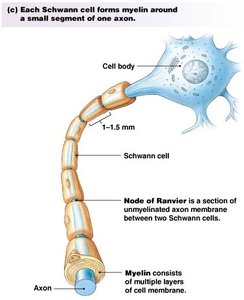
Peripheral Nerves
Peripheral nerves are bundles of axons surrounded by connective tissue layers (endoneurium, perineurium, epineurium). Schwann cells form myelin sheaths around axons, facilitating rapid signal conduction.
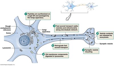
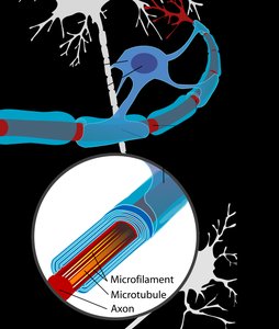
Axonal Transport
Axonal transport is essential for moving proteins, organelles, and other materials between the cell body and axon terminals. It occurs via two main mechanisms:
Fast Axonal Transport: Moves membrane-bound organelles (e.g., vesicles, mitochondria) at rates up to 400 mm/day (anterograde) and 200 mm/day (retrograde).
Slow Axonal Transport: Moves cytoplasmic proteins and cytoskeletal elements at rates up to 8 mm/day.

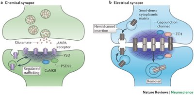
Synapses
Synapses are specialized junctions where neurons communicate with other neurons or effector cells. Most synapses are chemical, involving neurotransmitter release, while some are electrical, allowing direct ion flow through gap junctions.
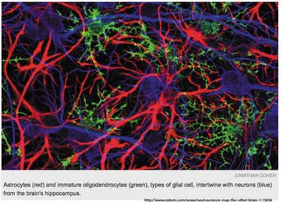
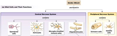
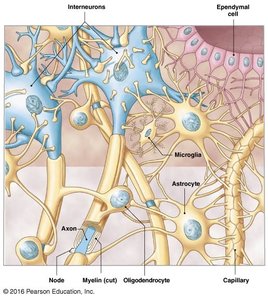
Glial Cells
Glial cells provide structural and metabolic support to neurons. Types include:
Astrocytes: Maintain extracellular environment, form the blood-brain barrier, and support synaptic function.
Oligodendrocytes (CNS) and Schwann Cells (PNS): Form myelin sheaths around axons.
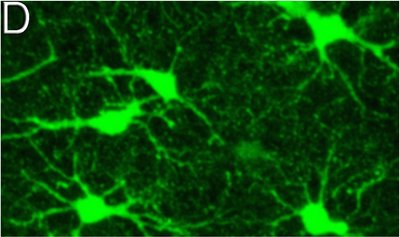
Microglia: Act as immune cells in the CNS.
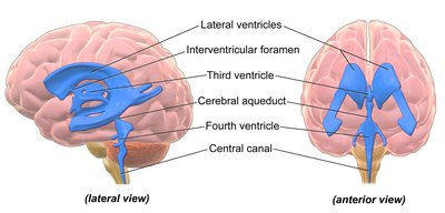
Ependymal Cells: Line fluid-filled cavities and help produce cerebrospinal fluid.
Satellite Cells (PNS): Support neuron cell bodies in ganglia.
Myelination and Demyelinating Diseases
Myelin is a multilayered phospholipid sheath that insulates axons, increasing the speed of electrical conduction (saltatory conduction). Oligodendrocytes myelinate CNS axons, while Schwann cells myelinate PNS axons. Demyelinating diseases, such as multiple sclerosis, disrupt this process, leading to neurological deficits.
Neuronal Injury and Regeneration
Peripheral neurons can regenerate if the cell body remains intact, guided by Schwann cells. In contrast, CNS repair is limited due to glial scar formation.
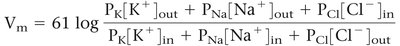
Electrical Signals in Neurons
Neurons and muscle cells are excitable, capable of generating and propagating electrical signals. The membrane potential is determined by the uneven distribution of ions and membrane permeability. The Nernst equation calculates the equilibrium potential for a single ion, while the Goldman-Hodgkin-Katz (GHK) equation predicts the overall membrane potential considering multiple ions.
Ion Channels and Membrane Potential
Resting Membrane Potential: Typically around -70 mV in neurons, mainly due to K+ permeability.
Gated Ion Channels: Include mechanically gated, chemically gated, and voltage-gated channels, each with specific triggers and properties.
Graded and Action Potentials
Graded Potentials: Variable-strength signals that decrease with distance; can be depolarizing (EPSP) or hyperpolarizing (IPSP).
Action Potentials: All-or-none electrical signals that propagate without loss of strength along the axon, initiated at the axon hillock if threshold is reached.
Summary Table: Major Types of Glial Cells
Glial Cell Type | Location | Main Function |
|---|---|---|
Astrocytes | CNS | Support, blood-brain barrier, regulate extracellular fluid |
Oligodendrocytes | CNS | Myelinate CNS axons |
Schwann Cells | PNS | Myelinate PNS axons |
Microglia | CNS | Immune defense |
Ependymal Cells | CNS | Line ventricles, produce cerebrospinal fluid |
Satellite Cells | PNS | Support neuron cell bodies in ganglia |
Key Equations
Nernst Equation: Calculates equilibrium potential for a single ion:
Goldman-Hodgkin-Katz (GHK) Equation: Predicts membrane potential considering multiple ions:
Ohm's Law: Describes current flow:
Additional info: This guide integrates foundational concepts from Ch. 8 (Neurons: Cellular and Network Properties) and related introductory physiology chapters, providing a comprehensive overview suitable for ANP college students.
