 Back
Neurophysiology and Muscle Physiology: ANP College Study Guide
Study Guide - Smart Notes
 Tailored notes based on your materials, expanded with key definitions, examples, and context.
Muscle Physiology
Sarcomere Structure and Function
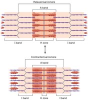
The sarcomere is the fundamental contractile unit of skeletal muscle, composed of thick and thin filaments. Understanding the arrangement and function of these filaments is essential for grasping muscle contraction mechanisms.
Thin Filament Proteins: The thin filament consists of actin, troponin, and tropomyosin. Myosin is not found in the thin filament; it is the main component of the thick filament.
Sarcomere Bands: The A band is the region of the sarcomere that contains the entire length of the thick filament (myosin), including areas where thick and thin filaments overlap.
Example: During muscle contraction, the sarcomere shortens, the I band and H zone decrease in width, but the A band remains unchanged.
Neuromuscular Junction
The neuromuscular junction is the synapse between a motor neuron and a skeletal muscle fiber, crucial for initiating muscle contraction.
Key Ion: Calcium (Ca2+) enters the axon terminal, triggering the release of the neurotransmitter acetylcholine into the synaptic cleft.
Function: Acetylcholine binds to receptors on the muscle cell membrane, leading to muscle activation.
Neurophysiology
Functions of the Nervous System
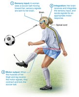
The nervous system is responsible for sensory input, integration, and motor output, enabling organisms to perceive and respond to their environment.
Sensory Input: Detects changes in the internal and external environment.
Integration: Processes and interprets sensory information to determine an appropriate response.
Motor Output: Activates effector organs (muscles and glands) to produce a response.
Divisions of the Nervous System
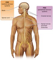
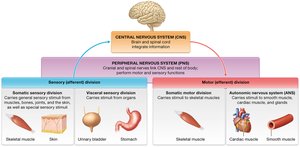
The nervous system is divided into the central nervous system (CNS) and peripheral nervous system (PNS), each with distinct structures and functions.
CNS: Composed of the brain and spinal cord; integrates information.
PNS: Includes cranial and spinal nerves; transmits information to and from the CNS.
Functional Divisions: Sensory (afferent) and motor (efferent) divisions, with the motor division further subdivided into somatic and autonomic systems.
Nervous Tissue
Nervous tissue consists of neurons and supporting glial cells, forming the structural and functional basis of the nervous system.
Neurons: Specialized cells for transmitting electrical signals.
Neuroglia: Support, protect, and nourish neurons.
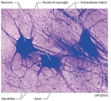
The Neuron
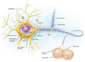
Neurons are the primary signaling cells of the nervous system, with distinct anatomical regions for receiving, conducting, and transmitting signals.
Anatomical Features: Dendrites (receive signals), cell body (integrates signals), axon (conducts signals), axon terminals (transmit signals).
Myelin Sheath: Insulates axons, increasing signal transmission speed.
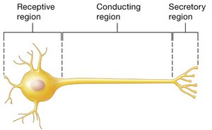
Functional Regions of Neurons
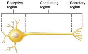
Neurons are organized into three functional regions: receptive, conducting, and secretory.
Receptive Region: Dendrites and cell body; receives input.
Conducting Region: Axon; transmits electrical impulses.
Secretory Region: Axon terminals; releases neurotransmitters.
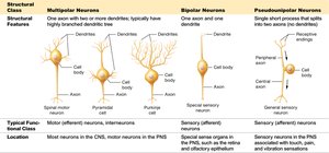
Classification of Neurons
Neurons are classified by function and structure, each with specific roles in the nervous system.
Sensory (Afferent) Neurons: Carry signals toward the CNS.
Motor (Efferent) Neurons: Carry signals away from the CNS to effectors.
Interneurons: Relay messages within the CNS.
Structural Types: Multipolar, bipolar, and pseudounipolar neurons.
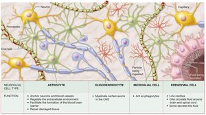
Neuroglia (Glial Cells)
Glial cells support neurons and are essential for nervous system function. They are more abundant than neurons and can divide throughout life.
CNS Glia: Astrocytes, oligodendrocytes, microglia, ependymal cells.
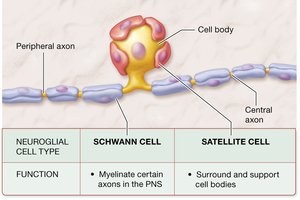
PNS Glia: Schwann cells, satellite cells.
Myelin Sheath Formation
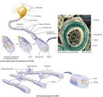
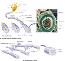
The myelin sheath is formed by oligodendrocytes in the CNS and Schwann cells in the PNS. It insulates axons and increases the speed of electrical signal transmission.
Composition: Repeating layers of glial cell plasma membrane, rich in lipids.
Function: Prevents ion leakage, ensures rapid signal conduction.
White Matter vs. Gray Matter
White matter consists of myelinated axons, while gray matter contains cell bodies and dendrites.
White Matter: Appears white due to myelin.
Gray Matter: Appears gray due to lack of myelin.
The Resting Membrane Potential
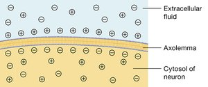
The resting membrane potential is the electrical charge difference across the plasma membrane of a neuron at rest, typically around -70 mV.
Cause: Leak channels allow potassium ions to leave the cell, making the inside more negative.
Polarization: The membrane has a positive and negative side.
Ion Channels in Electrically Excitable Cells
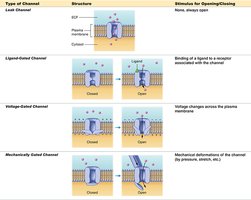
Ion channels regulate the flow of ions across the membrane, crucial for generating electrical signals.
Types: Leak channels (always open), ligand-gated channels (open in response to a chemical), voltage-gated channels (open in response to voltage changes), mechanically gated channels (open in response to physical deformation).
Depolarization and Hyperpolarization
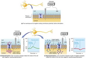
Depolarization and hyperpolarization are changes in membrane potential that affect neuron excitability.
Depolarization: Membrane potential becomes less negative, increasing likelihood of action potential.
Hyperpolarization: Membrane potential becomes more negative, decreasing likelihood of action potential.
The Action Potential
An action potential is a rapid, all-or-nothing electrical signal that travels along the axon of a neuron.
Trigger: Occurs when graded potentials reach threshold at the axon hillock.
Characteristics: All-or-nothing, travels entire axon, irreversible.
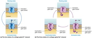
Voltage-Gated Channels (Sodium and Potassium)
Voltage-gated sodium and potassium channels are essential for the generation and propagation of action potentials.
Sodium Channels: Open rapidly during depolarization.
Potassium Channels: Open during repolarization.
Events of an Action Potential
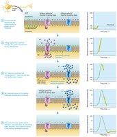
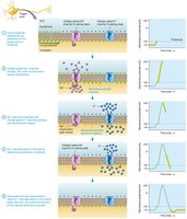
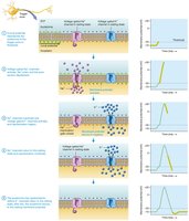
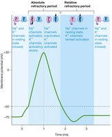
The action potential consists of several phases: depolarization, repolarization, and hyperpolarization.
Depolarization: Sodium channels open, Na+ enters the cell.
Repolarization: Potassium channels open, K+ leaves the cell.
Hyperpolarization: Membrane potential temporarily becomes more negative than resting.
The Sodium-Potassium Ion Pump
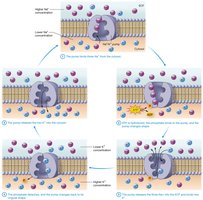
The sodium-potassium pump maintains the resting membrane potential by actively transporting sodium and potassium ions across the membrane.
Function: Moves 3 Na+ out and 2 K+ in per ATP molecule.
Equation:
The Refractory Period
The refractory period is the time during which a neuron cannot fire another action potential or can only fire with a strong stimulus.
Absolute Refractory Period: No additional action potential can be generated.
Relative Refractory Period: Only a strong stimulus can generate an action potential.
Local Potentials vs. Action Potentials
Local potentials are small, graded changes in membrane potential, while action potentials are large, all-or-nothing responses.
Local Potentials: Short distance, decremental, reversible, variable.
Action Potentials: Long distance, nondecremental, irreversible, stereotyped.
Action Potential Propagation
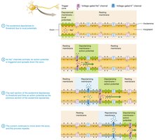
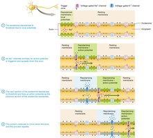
Action potentials propagate along the axon by triggering adjacent sections to depolarize, ensuring unidirectional flow due to refractory periods.
Mechanism: Self-propagating flow of charged particles.
Direction: Always forward, due to refractory period.
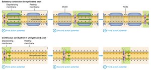
Saltatory vs. Continuous Conduction
Saltatory conduction occurs in myelinated axons, where action potentials jump between nodes of Ranvier, while continuous conduction occurs in unmyelinated axons.
Saltatory Conduction: Faster, action potential jumps from node to node.
Continuous Conduction: Slower, action potential renews at each segment.
Factors Influencing Conduction Speed
Conduction speed is affected by axon diameter and myelination.
Axon Diameter: Larger diameter = faster conduction.
Myelination: Myelinated axons conduct faster due to saltatory conduction.
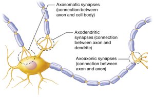
The Synapse
A synapse is the junction between a neuron and its target cell, enabling communication via electrical or chemical signals.
Types: Neuronal synapse (neuron-neuron), neuromuscular junction (neuron-muscle).
Components: Presynaptic neuron, postsynaptic neuron, synaptic cleft.
Electrical vs. Chemical Synapses
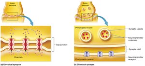
Synapses can be electrical (direct ion flow via gap junctions) or chemical (neurotransmitter-mediated).
Electrical Synapses: Bidirectional, instantaneous, coordinated activity.
Chemical Synapses: Unidirectional, slower, modifiable signal strength.
Chemical Synaptic Transmission
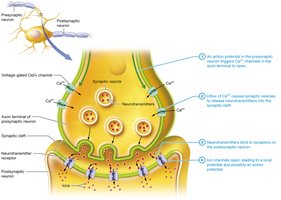
Chemical synaptic transmission involves the release of neurotransmitters from the presynaptic neuron, which bind to receptors on the postsynaptic neuron.
Steps: Action potential triggers Ca2+ influx, neurotransmitter release, receptor activation, postsynaptic potential generation.
Postsynaptic Potentials
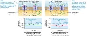
Postsynaptic potentials are local changes in membrane potential of the postsynaptic neuron, classified as excitatory (EPSP) or inhibitory (IPSP).
EPSP: Depolarizes membrane, increases likelihood of action potential.
IPSP: Hyperpolarizes membrane, decreases likelihood of action potential.
Neural Integration: Temporal and Spatial Summation
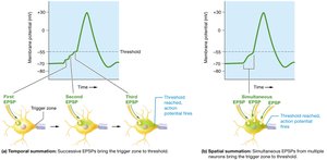
Neural integration is the process by which multiple postsynaptic potentials combine to influence the membrane potential at the trigger zone.
Temporal Summation: Repeated input from a single neuron.
Spatial Summation: Simultaneous input from multiple neurons.
Termination of Synaptic Transmission
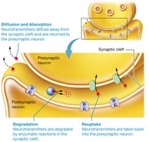
Synaptic transmission is terminated by diffusion, degradation, or reuptake of neurotransmitters.
Diffusion: Neurotransmitters diffuse away from the synaptic cleft.
Degradation: Enzymatic breakdown in the synaptic cleft.
Reuptake: Neurotransmitters are taken back into the presynaptic neuron.
Selected Neurotransmitters
Neurotransmitters are chemical messengers used by neurons to communicate.
Acetylcholine: Used at neuromuscular junctions.
Norepinephrine: Influences heart rate, blood pressure, digestion, attention.
Epinephrine: Similar to norepinephrine.
Dopamine: Modulates movement, emotion, motivation.
Serotonin: Modulates mood, attention, cognitive functions.
Additional info: These notes expand brief points into full academic explanations and include relevant images to reinforce key concepts. All content is structured for ANP college-level study and exam preparation.
