 Back
BackNeurophysiology and the Nervous System: Structure, Function, and Signaling
Study Guide - Smart Notes
 Tailored notes based on your materials, expanded with key definitions, examples, and context.
General Functions and Organization of the Nervous System
Overview and Functional Roles
The nervous system is the primary communication and control system of the body, responsible for collecting information, processing it, and initiating responses. It integrates sensory input and coordinates motor output to maintain homeostasis and regulate bodily functions.
Collecting Information: Receptors detect internal and external stimuli and send sensory signals to the brain and spinal cord.
Processing Information: The brain and spinal cord evaluate sensory input and determine appropriate responses.
Initiating Response: Motor output is relayed via nerves to effectors (muscles or glands).
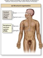
Structural Organization: CNS vs. PNS
The nervous system is divided into two main structural components:
Central Nervous System (CNS): Consists of the brain and spinal cord; responsible for integrating and processing information.
Peripheral Nervous System (PNS): Composed of nerves and ganglia; transmits signals between the CNS and the rest of the body.
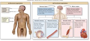
Functional Organization: Sensory vs. Motor
Sensory (Afferent) Division: Receives sensory information and transmits it to the CNS. Includes somatic (conscious perception) and visceral (unconscious perception) sensory systems.
Motor (Efferent) Division: Sends motor commands from the CNS to effectors. Includes somatic (voluntary control of skeletal muscles) and autonomic (involuntary control of smooth muscle, cardiac muscle, and glands) systems.
Brain Anatomy and Cerebral Organization
Major Brain Regions
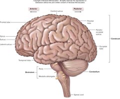
The brain is divided into four major regions, each with distinct functions:
Cerebrum: Responsible for complex intellectual functions, voluntary movement, and conscious perception.
Diencephalon: Integrates sensory and motor information.
Brainstem: Controls basic life functions.
Cerebellum: Coordinates movement and balance.
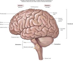
Cerebral Hemispheres and Lobes
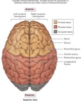
The cerebrum consists of two hemispheres, each divided into five lobes:
Frontal Lobe: Motor control, concentration, decision making, planning, personality.
Parietal Lobe: General sensory functions, evaluating shape and texture.
Temporal Lobe: Hearing and smell.
Occipital Lobe: Vision and visual memories.
Insula: Memory and sense of taste (not visible at the surface).
Nerves, Ganglia, and Neuron Structure
Structure of Nerves and Ganglia
Nerves are bundles of axons in the PNS, protected by three connective tissue wrappings:
Epineurium: Encloses the entire nerve.
Perineurium: Wraps each fascicle (bundle of axons).
Endoneurium: Wraps individual axons.
Ganglia are clusters of neuron cell bodies in the PNS. 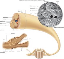
Neuron Structure and Functional Properties
Neurons are the structural units of the nervous system, exhibiting several key properties:
Excitability: Ability to respond to stimuli by changing membrane potential.
Conductivity: Ability to propagate electrical signals.
Secretion: Release of neurotransmitters to influence target cells.
Extreme Longevity: Neurons can live throughout a person's lifetime.
Amitotic: Most neurons lose the ability to divide after fetal development.
Parts of a Neuron
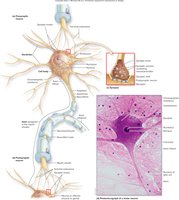
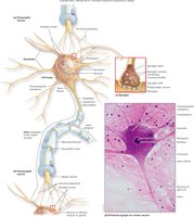
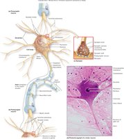
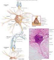
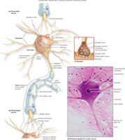
Cell Body (Soma): Contains nucleus and cytoplasm; initiates and receives graded potentials.
Dendrites: Short, branching processes that receive input and transfer it to the cell body.
Axon: Long process that conducts action potentials and releases neurotransmitters at synaptic knobs.
Neuron Transport
Axons transport materials via:
Anterograde Transport: Moves materials from cell body to synaptic knobs.
Retrograde Transport: Moves materials from axon to cell body for recycling.
Functional Classification of Neurons
Sensory (Afferent) Neurons: Conduct input from receptors to CNS; mostly unipolar.
Motor (Efferent) Neurons: Conduct output from CNS to effectors; multipolar.
Interneurons: Integrate information and communicate between sensory and motor neurons; multipolar and located in CNS.
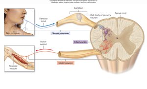
Synapses and Myelination
Chemical Synapses
Chemical synapses involve the release of neurotransmitters from the presynaptic neuron to the postsynaptic neuron across a synaptic cleft.
Synaptic Delay: Time required for neurotransmitter release, diffusion, and receptor binding.
Postsynaptic Potential: Graded potential initiated by neurotransmitter binding.
Myelination
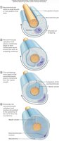
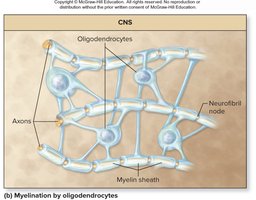
Myelination is the process of wrapping axons with myelin, which insulates and increases the speed of electrical conduction.
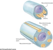
PNS: Neurolemmocytes (Schwann cells) myelinate axons; gaps are nodes of Ranvier.
CNS: Oligodendrocytes myelinate multiple axons; no neurilemma is formed.
Cell Communication and Electrical Activity
Ion Pumps and Channels
Neurons maintain concentration gradients of ions using pumps and channels:
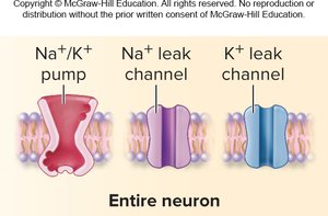
Leak Channels: Always open for continuous diffusion.
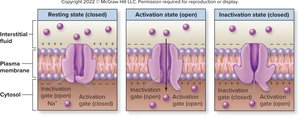
Chemically Gated Channels: Open in response to neurotransmitter binding.
Voltage-Gated Channels: Open in response to changes in membrane potential.
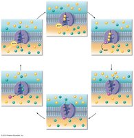
Pumps: Move ions against their concentration gradient using ATP (e.g., Na+/K+ pump).
Electrical Properties: Voltage, Current, and Resistance
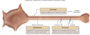
Voltage (V): Difference in electrical charge between two places; potential energy.
Current (I): Movement of charged particles across a barrier.
Resistance (R): Opposition to movement of charged particles.
Resting Membrane Potential (RMP)
Neurons at Rest
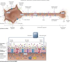
Ions are unevenly distributed across the plasma membrane due to pumps.
RMP is typically –70 mV, maintained by K+ diffusion and Na+/K+ pumps.
K+ diffusion is the primary factor in setting RMP; Na+ leak channels make RMP less negative.
Action Potential and Signal Propagation
All-or-None Law
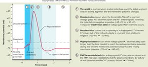
If threshold is reached, an action potential is generated and propagated without loss in intensity.
If threshold is not reached, no action potential occurs.
Events of an Action Potential
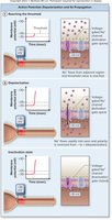
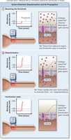
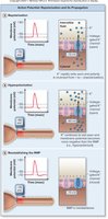
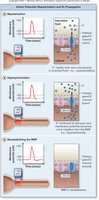
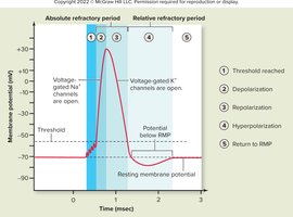
Depolarization: Na+ enters through voltage-gated channels, making membrane potential positive.
Repolarization: K+ exits through voltage-gated channels, returning membrane potential to negative.
Hyperpolarization: K+ channels stay open longer, making cell more negative than RMP.
Propagation: Action potential moves down the axon as voltage-gated channels open sequentially.
Refractory Periods
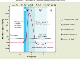
Absolute Refractory Period: No stimulus can initiate another action potential; ensures one-way propagation.
Relative Refractory Period: Another action potential is possible, but requires a stronger stimulus.
Propagation of Action Potential
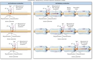
Continuous Conduction: Occurs in unmyelinated axons; slower.
Saltatory Conduction: Occurs in myelinated axons; faster, as action potential jumps between nodes of Ranvier.
Synaptic Transmission and Neurotransmitters
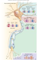
Transmissive Segment: Synaptic Knob Activity
Arrival of action potential opens voltage-gated Ca2+ channels.
Ca2+ triggers exocytosis of neurotransmitter vesicles.
Neurotransmitter binds to postsynaptic receptors, initiating a response.
Classification of Neurotransmitters
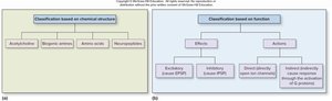
Neurotransmitters are classified by chemical structure and function:
Chemical Classes: Acetylcholine, biogenic amines, amino acids, neuropeptides.
Functional Classes: Excitatory or inhibitory; direct or indirect action.
Acetylcholine: Release and Removal
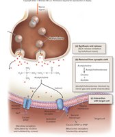
Synthesized from acetate and choline; stored in synaptic vesicles.
Released upon action potential arrival; binds to postsynaptic receptors.
Broken down by acetylcholinesterase; components recycled.
Neuromodulation
Facilitation and Inhibition
Neuromodulators alter the response of neurons:
Facilitation: Increases postsynaptic response by enhancing neurotransmitter release or receptor number.
Inhibition: Decreases postsynaptic response by reducing neurotransmitter release or receptor number.
Additional info: These mechanisms are essential for regulating neural circuits and adapting responses to changing physiological demands.
