 Back
BackOsseous Tissue and Bone Structure: Study Notes for Anatomy & Physiology
Study Guide - Smart Notes
 Tailored notes based on your materials, expanded with key definitions, examples, and context.
Osseous Tissue and Bone Structure
Skeletal System Functions
The skeletal system is essential for the structure, function, and maintenance of the human body. It provides support, stores minerals and lipids, produces blood cells, protects organs, and enables movement through leverage.
Support: Provides the structural framework for the body.
Storage of Minerals and Lipids: Bones store calcium, phosphorus, and energy reserves in yellow bone marrow.
Blood Cell Production: Red bone marrow produces red and white blood cells and other formed elements.
Protection: Surrounds and protects soft tissues and organs.
Leverage: Bones act as levers to change the magnitude and direction of muscle forces.
Bone Classifications
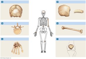
Bones are classified by their shapes and locations, which relate to their functions in the body.
Sutural Bones (Wormian Bones): Small, flat, irregular bones found between skull bones.
Irregular Bones: Complex shapes, such as vertebrae and pelvic bones.
Short Bones: Small, box-like bones, e.g., carpal and tarsal bones.
Flat Bones: Thin, parallel surfaces, e.g., skull roof, sternum, ribs, scapulae.
Long Bones: Long and slender, found in limbs, fingers, and toes.
Sesamoid Bones: Small, flat bones, e.g., patella; number varies among individuals.
Bone Markings and Structure
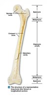
Bones have various markings that indicate attachments, articulations, and passageways for nerves and blood vessels. The structure of a typical long bone includes several distinct regions and tissues.
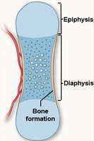
Diaphysis: The shaft of the bone.
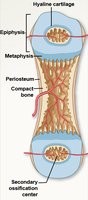
Epiphysis: The expanded end, mostly spongy bone.
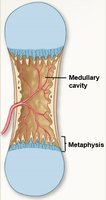
Metaphysis: Connects diaphysis and epiphysis.
Compact Bone: Dense outer layer providing strength.
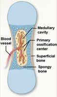
Medullary Cavity: Central space containing marrow.
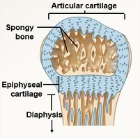
Spongy Bone: Lattice-like structure inside bones.
Common Bone Markings
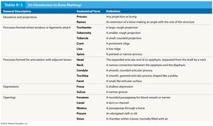
General Description | Anatomical Term | Definition |
|---|---|---|
Any projection or bump | Process | Any projection or bump |
Extension making an angle | Ramus | An extension of a bone making an angle with the rest of the structure |
Small, rounded projection | Tubercle | A small, rounded projection |
Large, rough projection | Tuberosity | A rough projection |
Prominent ridge | Crest | A prominent ridge |
Low ridge | Line | A low ridge |
Pointed process | Spine | A pointed process |
Expanded articular end | Head | The expanded articular end of an epiphysis, separated from the shaft by a neck |
Narrow connection | Neck | A narrow connection between the epiphysis and diaphysis |
Small, flat articular surface | Facet | A small, flat articular surface |
Rounded articular projection | Condyle | A smooth, rounded articular process |
Depression | Fossa | A shallow depression |
Passageway for nerves/vessels | Foramen | A rounded passageway for blood vessels or nerves |
Elongated cleft | Fissure | An elongated cleft |
Chamber within bone | Sinus | A chamber within a bone, normally filled with air |
Bone Composition
Bones are composed of four main cell types and a matrix of organic and inorganic materials.
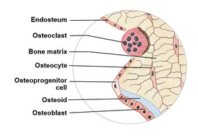
Osteocyte: Mature bone cell maintaining bone matrix.
Osteoprogenitor Cell: Stem cell producing new osteoblasts.
Osteoblast: Immature cell secreting bone matrix.
Osteoclast: Multinucleated cell dissolving bone matrix.
Bone matrix is about two-thirds calcium phosphate (forming hydroxyapatite crystals) and one-third collagen fibers, providing strength and flexibility.
Compact Bone Structure
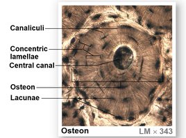
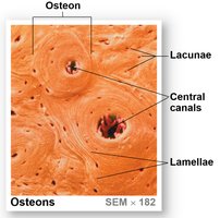
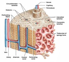
Compact bone is organized into osteons, the functional units of mature bone. Osteocytes are arranged in concentric lamellae around a central canal, which contains blood vessels and nerves.
Osteon: Structural unit of compact bone.
Central Canal: Contains blood vessels and nerves.
Perforating Canals: Connect central canals and supply deeper bone tissue.
Canaliculi: Tiny channels connecting lacunae and central canal.
Lacunae: Small spaces housing osteocytes.
Spongy Bone Structure
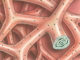
Spongy bone consists of a network of trabeculae, which are supporting bundles of fibers. Red bone marrow fills the spaces between trabeculae, producing blood cells and supplying nutrients. In some bones, yellow bone marrow (fat storage) is present.
Periosteum and Endosteum
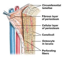
The periosteum is a double-layered membrane covering the bone's outer surface, except at joints. The endosteum is a thin cellular layer lining the medullary cavity and trabeculae of spongy bone.
Periosteum: Outer fibrous layer and inner cellular layer; isolates bone, provides blood supply, and aids in growth and repair.
Endosteum: Contains osteoblasts, osteoprogenitor cells, and osteoclasts; active in bone growth, repair, and remodeling.
Bone Growth and Ossification
Bone formation (ossification) begins about six weeks after fertilization and continues until about age 25. There are two main types of ossification: endochondral and intramembranous.
Ossification (Osteogenesis): Formation of bone tissue.
Calcification: Deposition of calcium salts in tissue.
Endochondral Ossification
Most bones develop from hyaline cartilage models through endochondral ossification. Bone formation begins at the primary ossification center and continues at secondary centers. Appositional growth increases bone diameter.
Epiphyseal Plate and Articular Cartilage
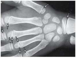
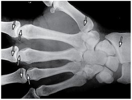
The epiphyseal plate (cartilage) separates the epiphysis and diaphysis during growth. When growth stops, the plate is replaced by bone, leaving an epiphyseal line. Articular cartilage remains at joint surfaces.
Intramembranous Ossification
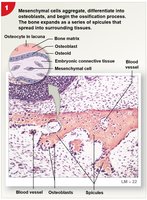
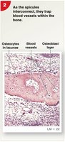
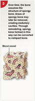
Intramembranous ossification occurs when osteoblasts differentiate within mesenchymal or fibrous connective tissue, forming dermal bones (e.g., skull, clavicle). Ossification centers form, and spicules trap osteoblasts and blood vessels.
Blood and Nerve Supply
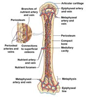
Bones receive blood from nutrient arteries and veins, metaphyseal vessels, and periosteal vessels. These vessels supply nutrients and oxygen to bone cells and remove waste. Bones also contain sensory nerves.
Bone Remodeling
Bone remodeling is a continuous process where old bone is replaced by new bone tissue. Osteoblasts and osteoclasts work together to maintain bone strength and mineral homeostasis. Remodeling rates vary by bone and region.
Hormonal Effects on Bone
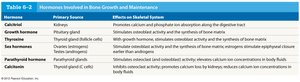
Bone growth and maintenance are regulated by several hormones, including calcitriol, growth hormone, thyroxine, sex hormones, parathyroid hormone, and calcitonin. These hormones influence bone formation, resorption, and mineral balance.
Hormone | Primary Source | Effect on Skeletal System |
|---|---|---|
Calcitriol | Kidneys | Promotes calcium and phosphate absorption in the digestive tract |
Growth hormone | Pituitary gland | Stimulates osteoblast activity and bone matrix synthesis |
Thyroxine | Thyroid gland | Stimulates cell metabolism and osteoblast activity |
Sex hormones | Ovaries/testes | Stimulate osteoblasts and produce bone growth |
Parathyroid hormone | Parathyroid glands | Stimulates osteoclast activity; increases blood calcium |
Calcitonin | Thyroid gland | Inhibits osteoclast activity; decreases blood calcium |
Ionic Composition of Bone
Bones serve as a major reservoir for minerals, especially calcium and phosphate. The skeleton contains 99% of the body's calcium, 80% of its carbonate, and significant amounts of other ions.
Calcium: 39% of bone mass
Phosphate: 17% of bone mass
Carbonate: 9.8% of bone mass
Other ions: Potassium, sodium, magnesium
About 67% of bone is inorganic (mainly hydroxyapatite), and 33% is organic (mainly collagen).
Blood Calcium Levels
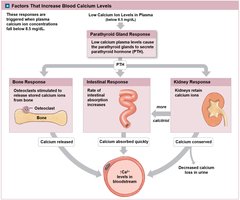
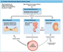
Blood calcium levels are tightly regulated by hormonal feedback involving the parathyroid and thyroid glands. Parathyroid hormone (PTH) increases blood calcium by stimulating osteoclasts, increasing intestinal absorption, and reducing renal excretion. Calcitonin decreases blood calcium by inhibiting osteoclasts and increasing renal excretion.
References: Martini, Frederic, Nath, Judi, and Bartholomew. Fundamentals of Anatomy and Physiology. Boston, MA: Benjamin Cummings, 2012. Print. Mescher, Anthony L., and Luiz Carlos Uchôa Junqueira. Junqueira's Basic Histology: Text and Atlas. Print.
