 Back
BackChapter 7 (Part IV): The Peripheral Nervous System (PNS) & the Autonomic Division
Study Guide - Smart Notes
 Tailored notes based on your materials, expanded with key definitions, examples, and context.
The Peripheral Nervous System (PNS) and Autonomic Division
Overview of the Nervous System
The nervous system is divided into the Central Nervous System (CNS) and the Peripheral Nervous System (PNS). The PNS consists of nerves and ganglia outside the CNS and is further divided into sensory and motor divisions. The motor division includes the somatic and autonomic divisions, with the autonomic division split into sympathetic and parasympathetic branches.
Sensory Receptors
Sensory receptors are specialized cells that detect changes in the environment (stimuli) and initiate nerve impulses. Sensation is the awareness of a stimulus, while perception is the interpretation of its meaning in the brain.
Mechanoreceptors: Respond to touch, pressure, vibration, stretch, and itch.
Thermoreceptors: Detect temperature changes.
Photoreceptors: Stimulated by light (e.g., retina).
Chemoreceptors: Respond to chemicals (e.g., smell, taste, blood chemistry).
Nociceptors: Sensitive to pain-causing stimuli.
Proprioceptors: Detect body position and movement.
Nerve Structure and Regeneration
Nerve Anatomy
Peripheral nerves are composed of bundles of axons (nerve fascicles), blood vessels, and connective tissues:
Endoneurium: Surrounds individual axons and their myelin sheaths.
Perineurium: Bundles groups of axons into fascicles.
Epineurium: Encloses the entire nerve.
Regeneration of Nerve Fibers
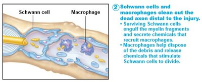
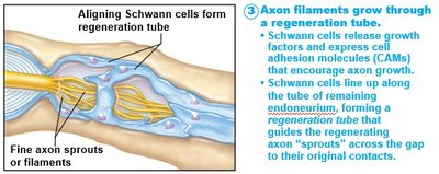
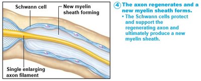
Unlike CNS neurons, PNS neurons can regenerate if the soma is intact. The process involves macrophages, Schwann cells, and axons.
Macrophages: Remove debris and stimulate Schwann cell division.
Schwann cells: Form a regeneration tube and secrete growth factors.
Axons: Regenerate and reconnect to their targets.
Cranial Nerves
Overview and Classification
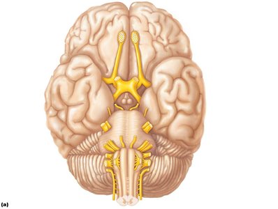
There are 12 pairs of cranial nerves, each identified by Roman numerals and names. They innervate the head and neck, except for the vagus nerve, which extends to thoracic and abdominal organs.
I Olfactory: Sensory; smell
II Optic: Sensory; vision
III Oculomotor: Motor; eye movement, pupil contraction
IV Trochlear: Motor; vertical eye movement
V Trigeminal: Both; facial sensation, mastication
VI Abducens: Motor; lateral eye movement
VII Facial: Both; taste, facial expression, salivation
VIII Vestibulocochlear: Sensory; hearing, equilibrium
IX Glossopharyngeal: Both; taste, swallowing, gag reflex
X Vagus: Both; parasympathetic functions, sensory from aorta, swallowing
XI Accessory: Motor; neck and shoulder movement
XII Hypoglossal: Motor; tongue movement, swallowing
Spinal Nerves
Classification and Function
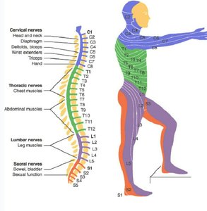
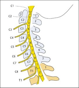
The spinal cord gives rise to 31 pairs of spinal nerves, classified by region:
8 cervical pairs (C1–C8)
12 thoracic pairs (T1–T12)
5 lumbar pairs (L1–L5)
5 sacral pairs (S1–S5)
1 coccygeal pair
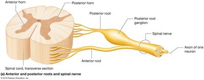
Each nerve pair innervates specific regions and functions of the body.
The Autonomic Nervous System (ANS)
Structure and Function
The ANS is part of the motor division of the PNS and regulates involuntary functions. It is divided into the sympathetic and parasympathetic divisions, both aiming to maintain homeostasis but through different mechanisms.
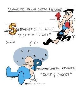
Sympathetic Division: Prepares the body for intense situations (fight, flight, freeze), moving away from homeostasis.
Parasympathetic Division: Returns the body to homeostasis, promoting rest and digest functions (SLUDDD: Salivation, Lacrimation, Urination, Digestion, Defecation, Decrease of heart rate).
Sympathetic vs. Parasympathetic Division
Both divisions have pre-synaptic and post-synaptic neurons, and their neurotransmitter effects depend on the receptor and target cell.
Sympathetic: Short pre-synaptic, long post-synaptic neurons; increases heart rate, blood pressure, bronchodilation, pupil dilation.
Parasympathetic: Long pre-synaptic, short post-synaptic neurons; decreases heart rate, promotes SLUDDD functions, bronchoconstriction, pupil constriction.
Homeostasis is the ultimate goal, with each division inhibiting the other to maintain balance.
Summary Table: Sympathetic vs. Parasympathetic Functions
Division | Main Functions | Neuron Structure | Response |
|---|---|---|---|
Sympathetic | Increase heart rate, blood pressure, bronchodilation, pupil dilation, glucose release | Short pre-synaptic, long post-synaptic | Fight, flight, freeze |
Parasympathetic | SLUDDD, decrease heart rate, bronchoconstriction, pupil constriction | Long pre-synaptic, short post-synaptic | Rest and digest |
Key Equations and Concepts
Action Potential:
Homeostasis: Maintenance of internal conditions despite external changes.
Additional info: Academic context was added to clarify the structure and function of the nervous system, cranial and spinal nerves, and the autonomic division. The included images directly reinforce nerve regeneration, cranial nerve locations, spinal nerve classification, and autonomic responses.
