 Back
BackPeripheral Nervous System and Reflex Activity: Structure, Function, and Clinical Relevance
Study Guide - Smart Notes
 Tailored notes based on your materials, expanded with key definitions, examples, and context.
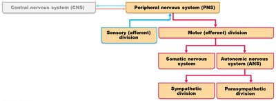
Peripheral Nervous System (PNS): Overview
General Features of the PNS
The Peripheral Nervous System (PNS) connects the central nervous system (CNS) to the rest of the body, providing essential communication links. It includes all neural structures outside the brain and spinal cord, such as sensory receptors, cranial nerves, peripheral nerves, ganglia, and motor endings.
Sensory receptors: Detect changes in the environment (stimuli).
Cranial nerves: Emerge from the brain, mainly serving the head and neck.
Spinal nerves: Arise from the spinal cord, serving the trunk and limbs.
Ganglia: Collections of neuron cell bodies outside the CNS.
Efferent motor endings: Activate effectors (muscles and glands).
Classification of Sensory Receptors
By Stimulus Type
Mechanoreceptors: Respond to touch, pressure, vibration, and stretch.
Thermoreceptors: Detect temperature changes.
Photoreceptors: Respond to light (e.g., in the retina).
Chemoreceptors: Detect chemicals (e.g., smell, taste, blood chemistry).
Nociceptors: Respond to pain-causing stimuli (e.g., extreme heat/cold, pressure, chemicals).
By Location
Exteroceptors: Respond to stimuli outside the body (e.g., skin, special senses).
Interoceptors (Visceroceptors): Respond to stimuli within the body (e.g., viscera, blood vessels).
Proprioceptors: Detect stretch in muscles, tendons, joints, and inform the brain about body position.
By Structural Complexity
Nonencapsulated (free nerve endings): Simple, widely distributed.
Encapsulated: Complex, with connective tissue coverings.
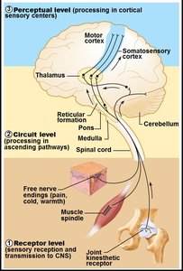
From Sensation to Perception
Neural Integration Levels
Sensation is the awareness of changes in the environment, while perception is the conscious interpretation of those stimuli. Neural integration occurs at three levels:
Receptor level: Sensory receptors detect stimuli and generate graded potentials.
Circuit level: Processing in ascending pathways to the brain.
Perceptual level: Processing in cortical sensory areas for conscious perception.
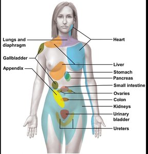
Visceral and Referred Pain
Key Elements
Visceral pain: Originates from internal organs, often described as aching or burning.
Referred pain: Pain perceived at a location other than the site of origin, due to convergence of visceral and somatic fibers in the same nerves (e.g., left arm pain during a heart attack).
Phantom limb pain: Pain perceived in an amputated limb, due to spinal cord and brain adaptation (hyperalgesia).
Peripheral Nerves and Ganglia
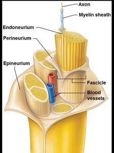
Structure of a Nerve
A nerve is a bundle of neuron fibers (axons) in the PNS, surrounded by connective tissue:
Endoneurium: Surrounds each axon.
Perineurium: Bundles groups of fibers into fascicles.
Epineurium: Encloses all fascicles to form the nerve.
Classification of Nerves
Mixed nerves: Contain both sensory and motor fibers.
Sensory (afferent) nerves: Carry impulses toward the CNS.
Motor (efferent) nerves: Carry impulses away from the CNS.
Peripheral nerves are classified as cranial or spinal nerves.
Classification of Ganglia
Sensory ganglia: Cell bodies of sensory neurons (e.g., dorsal root ganglia).
Autonomic ganglia: Cell bodies of autonomic motor neurons.
Cranial Nerves
Overview and Mnemonic
There are 12 pairs of cranial nerves, most serving the head and neck. The mnemonic "Oh, Oh, Oh, To Touch And Feel Very Green Vegetables, AH!" helps remember their order.
Olfactory (I)
Optic (II)
Oculomotor (III)
Trochlear (IV)
Trigeminal (V)
Abducens (VI)
Facial (VII)
Vestibulocochlear (VIII)
Glossopharyngeal (IX)
Vagus (X)
Accessory (XI)
Hypoglossal (XII)
Most cranial nerves are mixed, but some are purely sensory or motor. The vagus nerve is the only cranial nerve to extend beyond the head and neck.

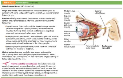
Example: Oculomotor Nerve (III)
Type: Motor
Function: Controls most eye movements, pupil constriction, and lens shape for focusing.
Origin: Ventral midbrain, passes through superior orbital fissure.
Spinal Nerves and Plexuses
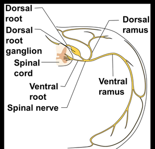
Spinal Nerves: Structure and Division
There are 31 pairs of spinal nerves, each formed by the combination of dorsal (sensory) and ventral (motor) roots. Spinal nerves quickly divide into dorsal and ventral rami, serving different body regions.

Nerve Plexuses
Except for T2–T12, ventral rami form interlacing nerve networks called plexuses:
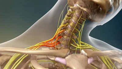
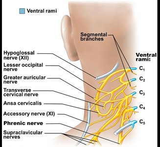
Cervical plexus (C1–C4): Innervates neck, ear, back of head, shoulders; phrenic nerve controls diaphragm.
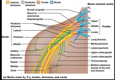
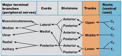
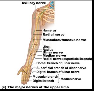
Brachial plexus (C5–T1): Innervates upper limb; major nerves include axillary, musculocutaneous, median, ulnar, and radial.
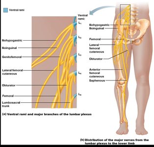
Lumbar plexus (L1–L4): Innervates thigh, abdominal wall; major nerves include femoral and obturator.
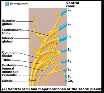
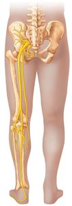
Sacral plexus (L4–S4): Innervates buttock, lower limb; major nerve is the sciatic (longest in the body).
Innervation of Thorax, Abdominal Wall, and Back
Ventral rami of T2–T12: Form intercostal nerves, supplying intercostal muscles, thorax, and abdominal wall.
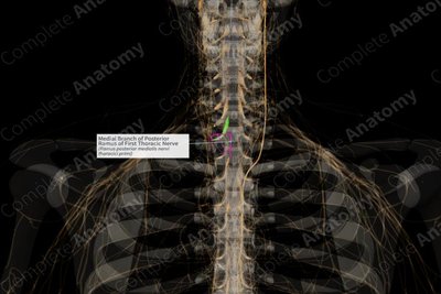
Dorsal rami: Innervate the posterior body trunk.
Dermatomes and Joint Innervation
Dermatomes
A dermatome is an area of skin innervated by the cutaneous branches of a single spinal nerve. Most dermatomes overlap, so damage to one nerve does not cause complete numbness.
Used clinically to assess spinal cord injuries.
Joint Innervation (Hilton's Law)
Any nerve serving a muscle that moves a joint also innervates the joint and the skin over it. For example, the knee joint is innervated by the femoral, sciatic, and obturator nerves.
Levels of Motor Control and Reflexes
Motor Control Hierarchy
Segmental level: Reflexes and automatic movements.
Projection level: Directs voluntary movements.
Precommand level: Cerebellum and basal nuclei plan and coordinate complex movements.
Reflexes and Reflex Arc
Inborn (intrinsic) reflexes: Rapid, involuntary, predictable responses (e.g., posture).
Learned (acquired) reflexes: Result from practice (e.g., driving).
The reflex arc consists of five components:
Receptor
Sensory neuron
Integration center
Motor neuron
Effector
Developmental Aspects and Disorders of the PNS
Development
Cranial and spinal nerves develop from neural crest cells.
With age, sensory receptors atrophy, muscle tone decreases, and reflexes slow.
Peripheral nerves remain viable unless damaged by trauma.
Disorders
Spinal cord trauma: Can cause flaccid or spastic paralysis.
Sciatica: Pain from irritation or injury to the sciatic nerve, causing pain, weakness, or numbness in the lower limb.
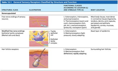
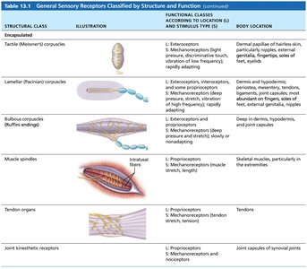
Summary Table: General Sensory Receptors
Structural Class | Illustration | Functional Class (Location & Stimulus) | Body Location |
|---|---|---|---|
Free nerve endings | Pain, temperature, pressure | Exteroceptors, interoceptors, proprioceptors; thermoreceptors, mechanoreceptors, nociceptors | Most body tissues, especially connective tissue and epithelia |
Merkel discs | Tactile epithelial complex | Exteroceptors; mechanoreceptors (light touch) | Basal layer of epidermis |
Hair follicle receptors | Hair deflection | Exteroceptors; mechanoreceptors (hair deflection) | Surrounding hair follicles |
Meissner's corpuscles | Tactile corpuscles | Exteroceptors; mechanoreceptors (light pressure, vibration) | Dermal papillae of hairless skin |
Lamellar corpuscles | Pacinian corpuscles | Exteroceptors, interoceptors, some proprioceptors; mechanoreceptors (deep pressure, vibration) | Dermis, hypodermis, periostea, mesentery, tendons, ligaments, joint capsules |
Muscle spindles | Intrafusal fibers | Proprioceptors; mechanoreceptors (muscle stretch, length) | Skeletal muscles, especially extremities |
Golgi tendon organs | Tendon organs | Proprioceptors; mechanoreceptors (tendon stretch, tension) | Tendons |
Joint kinesthetic receptors | Joint capsules | Proprioceptors; mechanoreceptors and nociceptors | Joint capsules of synovial joints |
