 Back
BackLecture - Ch 14 Peripheral Nervous System
Study Guide - Smart Notes
 Tailored notes based on your materials, expanded with key definitions, examples, and context.
Peripheral Nervous System (PNS)
Overview and Structural Organization
The Peripheral Nervous System (PNS) serves as the communication network linking the body to the Central Nervous System (CNS). It consists of all neural structures outside the brain and spinal cord, including sensory receptors, nerves, ganglia, and motor endings. The PNS is divided into sensory (afferent) and motor (efferent) divisions, with the motor division further subdivided into the somatic and autonomic nervous systems.
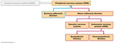
Sensory (Afferent) Division: Transmits sensory information from receptors to the CNS.
Motor (Efferent) Division: Transmits motor commands from the CNS to effectors (muscles and glands).
Somatic Nervous System: Controls voluntary movements of skeletal muscles.
Autonomic Nervous System (ANS): Regulates involuntary functions (e.g., heart rate, digestion) and is further divided into sympathetic and parasympathetic divisions.
Sensory Receptors and Sensation
Types and Classification of Sensory Receptors
Sensory receptors are specialized to detect changes in the environment (stimuli) and initiate nerve impulses. Sensation is the awareness of a stimulus, while perception is the interpretation of its meaning. Receptors are classified by stimulus type, location, and structural complexity.
By Stimulus Type:
Mechanoreceptors: Respond to touch, pressure, vibration, and stretch.
Thermoreceptors: Detect temperature changes.
Photoreceptors: Respond to light (e.g., retina).
Chemoreceptors: Detect chemicals (e.g., smell, taste, blood chemistry).
Nociceptors: Respond to pain-causing stimuli (e.g., extreme heat, pressure, chemicals).
Proprioceptors: Detect stretch in muscles, tendons, joints, and inform the brain of body position.
By Location:
Exteroceptors: Respond to stimuli outside the body (e.g., skin, special senses).
Interoceptors (Visceroceptors): Respond to stimuli within the body (e.g., internal organs, blood vessels).
Proprioceptors: Located in muscles, tendons, joints, and connective tissue coverings.
Perception of Pain
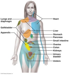
Visceral and Referred Pain
Visceral pain arises from stimulation of receptors in internal organs and is often felt as a vague, aching, or burning sensation. Referred pain occurs when pain from one region is perceived as coming from another, due to shared nerve pathways. For example, pain from a heart attack may be felt in the left arm.
Clinical Note: Long-lasting or intense pain can lead to hyperalgesia (pain amplification), chronic pain, and phantom limb pain. Early pain management is crucial to prevent these conditions.
Nerves and Associated Ganglia
Structure and Classification of Nerves
A nerve is a cordlike organ of the PNS, consisting of bundles of myelinated and unmyelinated axons enclosed by connective tissue. Nerves are classified as spinal or cranial based on their origin, and as sensory, motor, or mixed based on the direction of impulse transmission.
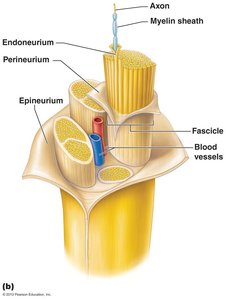
Connective Tissue Coverings:
Endoneurium: Surrounds individual axons and their myelin sheaths.
Perineurium: Bundles axons into fascicles.
Epineurium: Encloses all fascicles to form the nerve.
Types of Nerves:
Mixed nerves: Contain both sensory and motor fibers.
Sensory (afferent) nerves: Carry impulses toward the CNS.
Motor (efferent) nerves: Carry impulses away from the CNS.
Ganglia: Collections of neuron cell bodies in the PNS. Dorsal root ganglia contain sensory neuron cell bodies; autonomic ganglia contain motor neuron cell bodies.
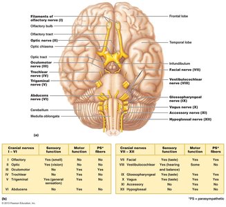
Cranial Nerves
Overview and Functions
There are 12 pairs of cranial nerves associated with the brain, each with specific sensory, motor, or mixed functions. Most originate from the brainstem, except the first two pairs. Cranial nerves are numbered I–XII from rostral to caudal.
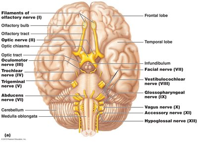
Mnemonic for Names: "On occasion, our trusty truck acts funny—very good vehicle anyhow."
Mnemonic for Functions: "Some say marry money, but my brother believes (it’s) bad business (to) marry money."
Summary Table: Cranial Nerve Functions
Cranial Nerve | Function | Type |
|---|---|---|
I. Olfactory | Smell | Sensory |
II. Optic | Vision | Sensory |
III. Oculomotor | Eye movement, pupil constriction | Motor |
IV. Trochlear | Eye movement (superior oblique) | Motor |
V. Trigeminal | Facial sensation, mastication | Mixed |
VI. Abducens | Eye movement (lateral rectus) | Motor |
VII. Facial | Facial expression, taste, salivation | Mixed |
VIII. Vestibulocochlear | Hearing, balance | Sensory |
IX. Glossopharyngeal | Taste, swallowing, salivation | Mixed |
X. Vagus | Autonomic control of viscera | Mixed |
XI. Accessory | Head and shoulder movement | Motor |
XII. Hypoglossal | Tongue movement | Motor |
Spinal Nerves and Plexuses
Spinal Nerve Structure and Distribution
There are 31 pairs of spinal nerves, all of which are mixed nerves. They are named according to their point of issue from the spinal cord and supply all body parts except the head and part of the neck. Each spinal nerve connects to the spinal cord via dorsal (sensory) and ventral (motor) roots, which join to form the nerve.
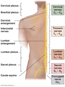
Cervical nerves: 8 pairs (C1–C8)
Thoracic nerves: 12 pairs (T1–T12)
Lumbar nerves: 5 pairs (L1–L5)
Sacral nerves: 5 pairs (S1–S5)
Coccygeal nerve: 1 pair (C0)
Nerve Plexuses
Nerve plexuses are interlacing networks of nerves formed by the ventral rami of spinal nerves (except T2–T12). The main plexuses are cervical, brachial, lumbar, and sacral, each serving specific body regions and providing redundancy in innervation.
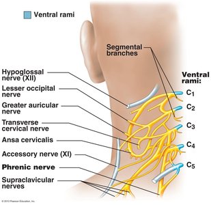
Cervical Plexus (C1–C4): Innervates skin and muscles of the neck, ear, back of head, and shoulders. The phrenic nerve (C3–C5) is the major motor and sensory nerve of the diaphragm.
Brachial Plexus (C5–T1): Innervates the upper limb. Major nerves include axillary, musculocutaneous, median, ulnar, and radial nerves.
Lumbar Plexus (L1–L4): Innervates the thigh, abdominal wall, and psoas muscle. Major nerves include femoral and obturator nerves.
Sacral Plexus (L4–S4): Serves the buttock, lower limb, pelvic structures, and perineum. The sciatic nerve is the largest and thickest nerve in the body.
Reflex Activity
Reflex Arcs and Types of Reflexes
A reflex is a rapid, involuntary response to a stimulus. Reflex arcs are the neural pathways that mediate reflexes and consist of five components: receptor, sensory neuron, integration center, motor neuron, and effector. Reflexes are classified as somatic (activating skeletal muscle) or autonomic (activating smooth/cardiac muscle or glands).
Inborn (intrinsic) reflexes: Rapid, predictable motor responses (e.g., posture maintenance).
Learned (acquired) reflexes: Result from practice or repetition (e.g., driving skills).
Developmental and Clinical Aspects
Development and Aging of the PNS
Spinal nerves branch from the developing spinal cord and neural crest cells, supplying both motor and sensory fibers to developing muscles. With age, sensory receptors atrophy, muscle tone decreases, and reflexes slow due to fewer synapses per neuron and slower central processing. Peripheral nerves remain viable unless damaged by trauma.
Clinical Correlations
Phantom limb pain: Pain felt in an amputated limb due to nerve pathway activity.
Carpal tunnel syndrome: Compression of the median nerve causing difficulty with thumb and finger movements.
Claw hand: Ulnar nerve injury causing characteristic hand deformity.
Wrist drop: Radial nerve injury resulting in inability to extend the hand at the wrist.
Sciatica: Pain radiating along the sciatic nerve, often due to disc herniation or injury.
Foot drop: Inability to dorsiflex the foot due to sciatic or peroneal nerve injury.
