 Back
Practical Review: Blood, Cardiovascular, and Lymphatic Systems
Study Guide - Smart Notes
 Tailored notes based on your materials, expanded with key definitions, examples, and context.
Blood: Components and Clinical Tests
Composition of Blood
Blood is a specialized connective tissue composed of plasma and formed elements. When whole blood is separated, it forms three distinct layers:
Plasma: The top layer, a straw-colored fluid, makes up about 55% of blood volume and contains water, proteins, electrolytes, and nutrients.
Buffy coat: The middle thin layer, composed of leukocytes (white blood cells) and platelets.
Erythrocytes: The bottom layer, consisting of red blood cells, accounts for about 45% of blood volume.
Formed elements are derived from hematopoietic stem cells in the bone marrow.
Characteristics and Functions of Formed Elements
Erythrocytes (Red Blood Cells): Biconcave, anucleate cells specialized for oxygen transport via hemoglobin.
Platelets: Small cell fragments involved in blood clotting.
Leukocytes (White Blood Cells): Immune cells classified as granulocytes or agranulocytes.
Granulocytes:
Neutrophils: Phagocytic cells, most abundant, respond to infection.
Eosinophils: Combat parasitic infections and modulate allergic responses.
Basophils: Release histamine, involved in inflammatory responses.
Agranulocytes:
Monocytes: Differentiate into macrophages, phagocytize pathogens.
Lymphocytes: Include B cells (antibody production) and T cells (cell-mediated immunity).
Clinical Blood Tests
Blood Typing System: Blood types are determined by antigens present on erythrocyte surfaces.
Antigens: A, B, Rh (D) are the main antigens involved.
Antibodies: Individuals produce antibodies against antigens not present on their own RBCs.
Agglutination is the clumping of cells due to antibody-antigen interaction, used to determine blood types. Types of Anemia:
Iron-deficiency anemia
Pernicious anemia
Hemorrhagic anemia
Hemolytic anemia
Aplastic anemia
Heart Anatomy and Physiology
Heart Structure and Location
The heart is located in the mediastinum, surrounded by serous membranes (pericardium), and composed of cardiac muscle tissue. The heart wall consists of three layers:
Epicardium: Outer layer
Myocardium: Middle, muscular layer
Endocardium: Inner layer
Heart Chambers, Valves, and Great Vessels
Chambers: Right and left atria, right and left ventricles
Valves: Tricuspid, bicuspid (mitral), pulmonary, aortic
Great vessels: Superior/inferior vena cava, pulmonary arteries/veins, aorta
Blood Flow Direction:
Deoxygenated blood enters right atrium → right ventricle → pulmonary arteries → lungs
Oxygenated blood returns via pulmonary veins → left atrium → left ventricle → aorta → systemic circulation
Cardiovascular Physiology
Blood Pressure
Systolic pressure: Pressure during ventricular contraction
Diastolic pressure: Pressure during ventricular relaxation
Conduction System of the Heart
Sinoatrial (SA) node: Pacemaker, initiates electrical impulse
Atrioventricular (AV) node: Delays impulse, allows atrial contraction
Bundle of His, bundle branches, Purkinje fibers: Distribute impulse to ventricles
Electrocardiogram (EKG)
P wave: Atrial depolarization
QRS complex: Ventricular depolarization
T wave: Ventricular repolarization
Types of Arrhythmias
Bradycardia: Slow heart rate
Tachycardia: Fast heart rate
Atrial fibrillation (A-fib): Irregular atrial rhythm
Ventricular fibrillation (V-fib): Disorganized ventricular rhythm
Blood Vessels
Types and Structure of Blood Vessels
There are three basic types of blood vessels:
Arteries: Carry blood away from the heart; thick muscular layer, highly folded endothelium
Veins: Carry blood toward the heart; smooth endothelium, valves, less smooth muscle
Capillaries: Site of exchange; simple squamous epithelium
Layers of Vessel Walls:
Tunica intima: Inner layer
Tunica media: Middle, muscular layer
Tunica externa: Outer layer
Great Vessels of Pulmonary and Systemic Circuits
Pulmonary circuit: Pulmonary arteries and veins
Systemic circuit: Aorta, vena cavae, and branches
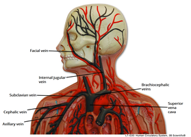
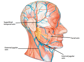
Systemic Vessels: Head and Neck
The head and neck contain major arteries and veins supplying and draining blood.
Common carotid artery
Temporal artery
Occipital artery
Facial artery
External and internal jugular veins
Systemic Vessels: Arm
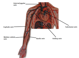
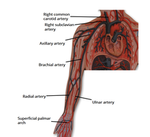
The arm is supplied by branches from the subclavian artery and drained by corresponding veins.
Arteries: Axillary, brachial, radial, ulnar, palmar arch
Veins: Dorsal venous network, basilic (medial), cephalic (lateral), axillary, subclavian
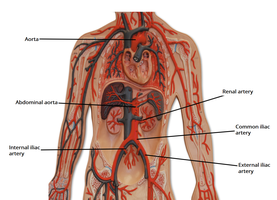
Systemic Vessels: Thorax and Abdomen
The thorax and abdomen contain the aorta and its branches, as well as veins associated with organs.
Aorta: Ascending, arch, descending (thoracic and abdominal)
Celiac trunk: Supplies upper abdominal viscera
Common iliac, sacral arteries
Hepatic, splenic, renal arteries and veins
Systemic Vessels: Leg
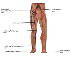
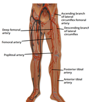
The leg is supplied by femoral and tibial arteries and drained by saphenous and other veins.
Arteries: Femoral, deep femoral, popliteal, anterior and posterior tibial
Veins: Great and small saphenous, dorsal venous arch
Lymphatic System
Lymphatic Organs and Functions
The lymphatic system is essential for immune defense and fluid balance.
Lymph nodes: Filter lymph, house immune cells
Spleen: Filters blood, recycles RBCs, stores platelets
Thymus gland: Site of T cell maturation
Organization of Lymphatic Vessels
Lymph flows through a hierarchical network:
Lymphatic capillaries
Lymphatic vessels
Lymphatic trunks
Lymphatic ducts (right lymphatic duct and thoracic duct)
Right and left subclavian veins
Inflammatory Response
The inflammatory response is a protective reaction to injury or infection.
Pain
Redness
Swelling
Heat
Steps:
Release of inflammatory mediators
Vasodilation and increased permeability
Recruitment of immune cells
Resolution and tissue repair
Example: A cut on the skin triggers inflammation, resulting in redness, swelling, heat, and pain as immune cells respond to pathogens.
Additional info: Academic context was added to clarify vessel structure, blood cell function, and lymphatic organization.
