 Back
BackRenal Anatomy and Physiology: Structure and Function of the Kidney and Nephron
Study Guide - Smart Notes
 Tailored notes based on your materials, expanded with key definitions, examples, and context.
The Urinary System
Overview and Importance
The urinary system is essential for maintaining homeostasis by filtering blood, removing waste products, and regulating fluid and electrolyte balance. Understanding normal kidney function is crucial for recognizing and treating kidney pathophysiology.
Major Organs: Kidneys, ureters, bladder, and urethra.
Primary Functions of Kidneys:
Excretion of metabolic waste products (e.g., urea, creatinine).
Elimination of foreign compounds (e.g., drugs).
Regulation of long-term acid-base balance.
Long-term control of blood pressure.
Processing approximately 180 L of plasma per day.
Urine Formation: Filtrate formed in kidneys travels via ureters to the bladder for storage and is excreted through the urethra.

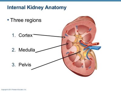
Kidney Structure
Gross Anatomy
The kidneys are paired, bean-shaped organs located on the posterior abdominal wall, outside the peritoneal cavity. Each kidney contains 800,000 to 1.5 million nephrons, the functional units responsible for filtration and urine formation.
Zones of the Kidney:
Renal Cortex: The outer layer, containing glomeruli and convoluted tubules.
Renal Medulla: The inner region, organized into pyramids, primarily containing loops of Henle and collecting ducts.
Renal Pelvis: The funnel-shaped structure that collects urine and channels it into the ureter.
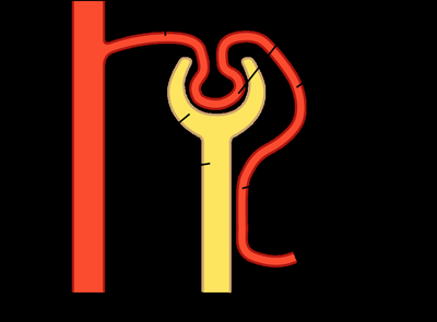
Nephron Structure and Function
Overview of the Nephron
The nephron is the microscopic structural and functional unit of the kidney. Each nephron consists of a renal corpuscle (glomerulus and Bowman’s capsule) and a renal tubule (proximal tubule, loop of Henle, distal tubule, and collecting duct).
Vascular Components:
Renal artery: Supplies blood to the kidney.
Afferent arteriole: Brings blood to the glomerulus.
Glomerulus: Ball of capillaries where filtration occurs.
Efferent arteriole: Carries blood away from the glomerulus.
Peritubular capillaries: Surround the tubules, facilitating exchange of substances between blood and tubular fluid.
Renal vein: Returns filtered blood to systemic circulation.
Tubular Components:
Bowman’s capsule: Surrounds the glomerulus and collects filtrate.
Proximal tubule: Major site of reabsorption of water, ions, and nutrients.
Loop of Henle: Establishes osmotic gradient; descending limb permeable to water, ascending limb permeable to sodium.
Distal tubule and collecting duct: Fine-tune reabsorption and secretion, regulated by hormones (e.g., aldosterone, ADH).
Renal Blood Flow and Filtration
Glomerular Filtration Rate (GFR)
GFR is the volume of plasma filtered by the glomeruli per minute and is a key indicator of kidney function. In healthy adults, GFR is about 125 mL/min (180 L/day), meaning the entire plasma volume is filtered approximately 60 times per day.
Filtration Fraction (FF): The fraction of renal plasma flow filtered into the glomerulus per pass.
Formula:
Example: (20%)
99% of filtrate is reabsorbed; less than 1% is excreted as urine.
Forces Driving Glomerular Filtration
Filtration in the glomerulus is determined by the balance of hydrostatic and osmotic pressures:
Forces Favoring Filtration:
Glomerular capillary hydrostatic pressure (from the heart)
Forces Opposing Filtration:
Hydrostatic pressure in Bowman’s capsule
Osmotic pressure due to plasma proteins (plasma oncotic pressure)
Net filtration pressure determines the GFR. Changes in these forces can alter GFR, especially in pathological conditions (e.g., burns, urinary tract obstruction, dehydration).
Glomerular Filtration Barrier
Structure and Function
The glomerular filtration barrier is a highly selective structure that allows passage of water and small solutes but restricts proteins and cells.
Three Layers:
Fenestrated endothelial cells
Negatively charged basement membrane
Podocytes with slit diaphragms
Filterability: Small molecules (<3 nm or 7000 MW) pass freely; large proteins (>7-9 nm or 70000 MW) are blocked, especially due to negative charge.
Filtrate in Bowman’s capsule is nearly protein-free.
Damage to the filtration barrier (e.g., in diabetes) can cause proteinuria (e.g., albuminuria), indicating kidney disease.
Regulation of Glomerular Filtration Rate (GFR)
Autoregulation Mechanisms
The kidneys maintain a relatively constant GFR despite fluctuations in blood pressure through autoregulation. This prevents imbalances in fluid, electrolyte, and waste excretion and protects the filtration barrier from damage.
Myogenic Mechanism: Arteriolar smooth muscle contracts in response to stretch (increased blood pressure), reducing afferent arteriole diameter and stabilizing GFR.
Tubuloglomerular Feedback: Macula densa cells in the distal tubule sense NaCl concentration. Increased NaCl (from increased GFR) signals afferent arteriole constriction, reducing GFR. Juxtaglomerular cells release renin, influencing GFR via the renin-angiotensin system.
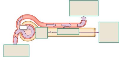
Tubular Reabsorption and Secretion
Processes in the Nephron
After filtration, the nephron modifies the filtrate through reabsorption and secretion:
Reabsorption: Movement of substances from the tubule back into the blood (peritubular capillaries). Most water, ions, and nutrients are reabsorbed.
Secretion: Movement of substances from blood into the tubule for excretion (e.g., drugs, excess K+, H+).
Excretion: The final urine output, calculated as:
Reabsorption is selective and regulated by hormones (e.g., aldosterone for sodium, ADH for water). The transport maximum (Tm) limits the rate of active reabsorption (e.g., glucose reabsorption is saturated in diabetes, leading to glucosuria).
Summary Table: Main Functions and Components of the Nephron
Component | Main Function | Key Features |
|---|---|---|
Bowman’s Capsule | Filtration | Collects filtrate from glomerulus |
Proximal Tubule | Reabsorption | Major site for water, ion, and nutrient reabsorption |
Loop of Henle | Concentration of urine | Descending limb: water reabsorption; Ascending limb: sodium reabsorption |
Distal Tubule & Collecting Duct | Fine-tuning reabsorption/secretion | Regulated by aldosterone (Na+) and ADH (water) |
Key Equations
Filtration Fraction:
Excretion Rate:
Creatinine Clearance (estimate of GFR):
Clinical Relevance
GFR declines in kidney disease; eGFR is used clinically to assess renal function.
Proteinuria (e.g., albuminuria) is a marker of glomerular barrier dysfunction.
Chronic kidney disease (CKD) leads to progressive loss of function, requiring dialysis or transplantation in end-stage disease.
