Back
BackRenal System: Anatomy and Physiology Study Notes
Study Guide - Smart Notes
 Tailored notes based on your materials, expanded with key definitions, examples, and context.
Renal System Overview
Importance of the Kidneys
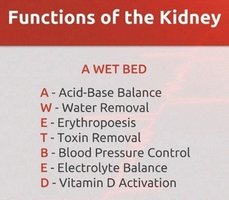
The kidneys are vital organs responsible for maintaining homeostasis by regulating fluid, electrolyte, and acid-base balance, as well as removing metabolic wastes. They also play key roles in hormone production and vitamin D activation.
Salt and water balance: Maintains osmotic equilibrium and blood volume.
Regulation of blood ion concentrations: Controls levels of sodium, potassium, calcium, and other ions.
Acid-base balance: Adjusts pH by excreting hydrogen ions and reabsorbing bicarbonate.
Excretion of metabolic wastes, drugs, and toxins: Removes urea, creatinine, and other waste products.
Production of erythropoietin (EPO): Stimulates red blood cell production.
Production of renin: Regulates blood pressure.
Activation of vitamin D: Converts vitamin D to its active form for calcium absorption.
Anatomy of the Urinary System
Gross Anatomy of the Kidneys
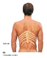
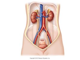
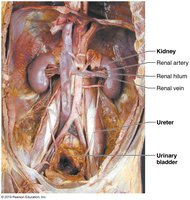
The kidneys are bean-shaped, retroperitoneal organs located in the superior lumbar region, protected partially by the rib cage. The right kidney is slightly lower due to the liver. Each kidney weighs about 150 g and measures approximately 12 x 6 x 3 cm. The adrenal glands sit atop each kidney.
Location: From the 12th thoracic vertebra to the 3rd lumbar vertebra.
Protection: Rib cage and perirenal fat capsule.
Renal hilum: Medial indentation where vessels and nerves enter/exit.
Supportive Tissue Layers
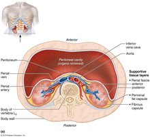
Three layers of supportive tissue surround the kidneys:
Renal (fibrous) capsule: Adheres directly to the kidney surface, providing a strong barrier.
Perirenal fat capsule: Cushions and helps hold the kidney in place.
Renal fascia: Dense connective tissue that anchors the kidney and adrenal gland.
Internal Anatomy of the Kidney
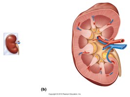
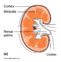
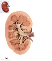
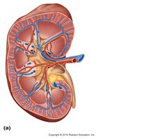
The kidney consists of three main regions: cortex, medulla, and pelvis. The cortex is the site of filtration, the medulla contains renal pyramids, and the pelvis collects urine before it enters the ureter.
Cortex: Site of glomerular filtration.
Medulla: Contains renal pyramids, which appear striped due to parallel arrangement of tubules and blood vessels.
Renal pelvis: Funnel-shaped tube continuous with the ureter; collects urine from calyces.
Calyces: Minor calyces enclose papillae of pyramids; major calyces collect urine from minor calyces.
Peristalsis: Smooth muscle in walls of calyces, pelvis, and ureter propels urine.
Blood and Nerve Supply
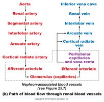
The kidneys receive about 25% of total systemic cardiac output (~1.2 L/min). Blood enters via the renal arteries, passes through a series of branches, and exits via the renal veins. The renal plexus, primarily composed of sympathetic fibers, regulates blood flow by adjusting arteriole diameters.
Renal artery → segmental artery → interlobar artery → arcuate artery → cortical radiate artery → afferent arteriole → glomerulus → efferent arteriole → peritubular capillaries/vasa recta → cortical radiate vein → arcuate vein → interlobar vein → renal vein
Nephron Structure and Types
Nephron Anatomy
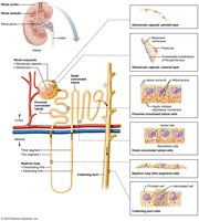
The nephron is the structural and functional unit of the kidney, responsible for filtering blood and forming urine. Each kidney contains about one million nephrons and thousands of collecting ducts.
Renal corpuscle: Includes the glomerulus and Bowman's capsule.
Renal tubule: Composed of the proximal convoluted tubule, nephron loop (loop of Henle), distal convoluted tubule, and collecting duct.
Collecting duct: Contains principal cells (salt and water balance) and intercalated cells (acid-base balance).
Types of Nephrons
There are two main types of nephrons:
Cortical nephrons (85%): Located mostly in the cortex; responsible for most filtration.
Juxtamedullary nephrons (15%): Located near the medulla; important for concentrating urine.
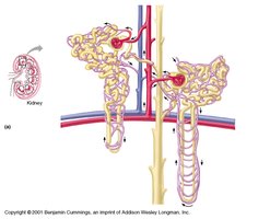
Microcirculation of the Nephron
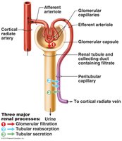
The glomerulus is specialized for filtration and is both fed and drained by arterioles. The afferent arteriole has a larger diameter, creating high pressure for filtration. Peritubular capillaries arise from efferent arterioles and drain into renal venules. The vasa recta is found in juxtamedullary nephrons and is essential for urine concentration.
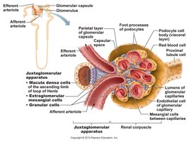
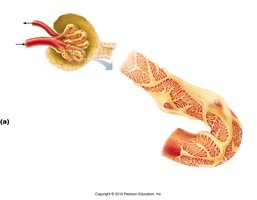
Juxtaglomerular Complex (JGC)
Structural Organization
The JGC is located at the junction of the early distal convoluted tubule and afferent/efferent arterioles. It regulates renal function and blood pressure.
Granular (JG) cells: Mechanoreceptors in arteriole walls; secrete renin.
Macula densa cells: Chemo- or osmoreceptors in tubule wall; monitor filtrate and adjust glomerular filtration rate (GFR).
Functions: Regulates filtrate formation and systemic blood pressure.
Micturition Pathways
Male vs. Female Urinary Tract
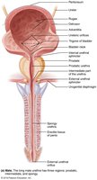
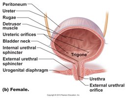
The micturition pathway differs between males and females, primarily in the length and structure of the urethra. The trigone is a clinically important area of the bladder, formed by the openings of the ureters and urethra.
Internal urethral sphincter: Formed by smooth muscle at the bladder neck.
External urethral sphincter: Formed by skeletal muscle in the urogenital diaphragm.
Bladder capacity: Moderately full bladder holds ~500 ml; can expand to ~1 L.
Renal Physiology
Nephron Functions
The kidneys filter several liters of fluid from the bloodstream daily, removing toxins, metabolic wastes, and excess ions while returning needed materials to the blood.
Primary functions: Excretion and homeostasis.
Additional functions: Acid-base balance, water removal, erythropoiesis, toxin removal, blood pressure control, electrolyte balance, vitamin D activation.
Processes of Urine Formation
Three major processes occur in the nephron:
Glomerular filtration: Plasma minus proteins is filtered into the renal tubules.
Tubular reabsorption: Nutrients, essential ions, and most water are reabsorbed into the bloodstream.
Tubular secretion: Additional wastes and excess ions are secreted into the tubule.
Filtrate is the fluid entering the tubules; urine is the final product after reabsorption and secretion.
Immunology Context (Relevant to Renal System)
Antibody Types and Functions
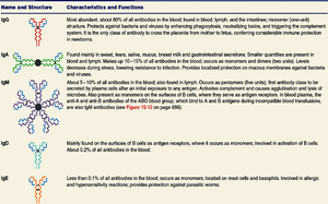
Antibodies are critical components of the immune system, with IgA being the main antibody found in secretions such as saliva and breastmilk. This is relevant to renal physiology due to the kidney's role in filtering blood and immune components.
Name and Structure | Characteristics and Functions |
|---|---|
IgG | Most abundant; protects against bacteria and viruses; crosses placenta. |
IgA | Found in secretions; protects mucosal surfaces. |
IgM | First antibody produced; activates complement. |
IgD | Found on B cells; involved in activation. |
IgE | Involved in allergic reactions; protects against parasites. |
Active vs. Passive Immunity
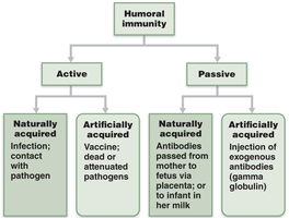
Humoral immunity can be acquired actively (through infection or vaccination) or passively (through maternal antibodies or injection of exogenous antibodies). Both mechanisms involve antibodies and are part of adaptive immunity.
T-Cell Maturation and Negative Selection
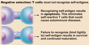
During T-cell maturation, recognition of self-antigen results in apoptosis, preventing autoimmune diseases. Failure to recognize self-antigen allows survival and continued maturation.