 Back
BackSensory Physiology and Autonomic Motor Control: Mini-Textbook Study Notes
Study Guide - Smart Notes
 Tailored notes based on your materials, expanded with key definitions, examples, and context.
Ch. 10 Sensory Physiology
General Properties of Sensory Systems
Sensory systems allow the body to detect and interpret information from both the external and internal environments. Sensory neurons possess specialized transducers (receptors) that convert physical stimuli into intracellular signals, typically changes in membrane potential. These signals are then processed by the nervous system to generate appropriate responses.
Transduction: The process by which a receptor converts a physical stimulus (e.g., light, pressure, chemicals) into an electrical signal.
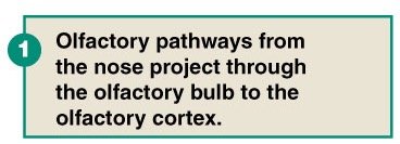
Special Senses: Vision, hearing, taste, smell, and equilibrium. Most are mediated by non-neuronal cells except olfaction.
Somatic Senses: Touch, temperature, pain, itch, and proprioception (sense of body position).
Receptor Types: Chemoreceptors, mechanoreceptors, thermoreceptors, nociceptors, and proprioceptors.
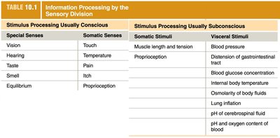
Conscious and Subconscious Sensory Processing
Sensory information can be processed consciously or subconsciously, depending on the type and location of the stimulus.
Stimulus Processing Usually Conscious | Stimulus Processing Usually Subconscious |
|---|---|
Special Senses Vision Hearing Taste Smell Equilibrium | Somatic Stimuli Muscle length and tension Proprioception |
Somatic Senses Touch Temperature Pain Itch Proprioception | Visceral Stimuli Blood pressure Distension of gastrointestinal tract Blood glucose concentration Internal body temperature Osmolarity of body fluids Lung inflation pH of cerebrospinal fluid pH and oxygen content of blood |
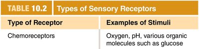
Types of Sensory Receptors
Sensory receptors are specialized to detect specific types of stimuli:
Type of Receptor | Examples of Stimuli |
|---|---|
Chemoreceptors | Oxygen, pH, various organic molecules such as glucose |
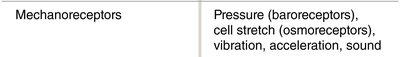
Mechanoreceptors | Pressure (baroreceptors), cell stretch (osmoreceptors), vibration, acceleration, sound |
Thermoreceptors | Varying degrees of heat |

Receptor Potentials and Action Potentials
Physical stimuli are transduced into receptor potentials, which are graded changes in membrane potential. If the receptor potential reaches threshold, it triggers action potentials in the sensory neuron, allowing the signal to be transmitted to the central nervous system (CNS).
Receptive Fields
A receptive field is the specific physical area where a stimulus will activate a particular sensory neuron. The size and overlap of receptive fields influence the precision of sensory perception.
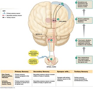
Somatosensory Pathways and CNS Integration
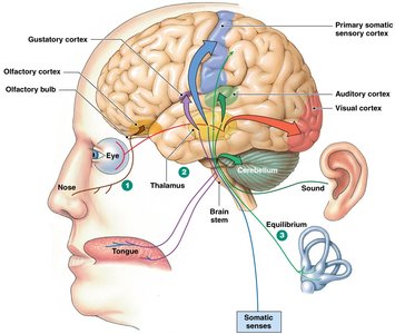
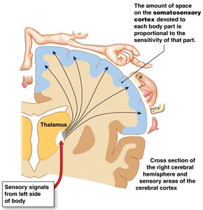
Somatosensory neurons carry information from the body to the CNS. Most sensory information is routed through the thalamus, which acts as a relay station. Special senses have dedicated cortical regions, while somatic senses are integrated in the primary somatosensory cortex. Visceral sensory information is integrated in the brainstem and spinal cord.
Coding and Processing of Sensory Information
The CNS distinguishes between different sensations by analyzing four main properties of a stimulus:
Modality: The type of stimulus, determined by the receptor and the pathway to the brain.
Location: Determined by which receptive fields are activated and where the pathways terminate in the brain.
Intensity: Determined by the number of receptors activated (population coding) and the frequency of action potentials (frequency coding).
Duration: Determined by how long action potentials are generated and by receptor adaptation.
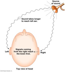
Sound Localization
Sound localization depends on the timing differences in which sound reaches each ear and is processed by the auditory cortex. Lateral inhibition can enhance the accuracy of localization by inhibiting neighboring neurons, sharpening the perception of stimulus location.
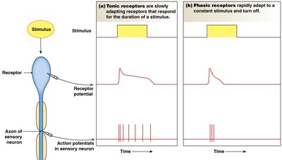
Receptor Adaptation
Receptors can adapt to constant stimuli in different ways:
Tonic receptors: Slowly adapting receptors that respond for the duration of a stimulus.
Phasic receptors: Rapidly adapt to a constant stimulus and turn off.
Sensory Pathway Specificity
Each sensory pathway is specific for a particular type of stimulus and projects to a specific region of the cerebral cortex. This allows the brain to identify the origin and nature of each incoming signal.
Ch. 11 Efferent Division: Autonomic and Somatic Motor Control
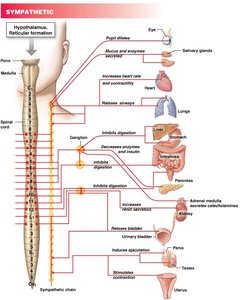
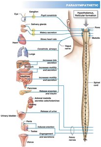
Autonomic Nervous System (ANS)
The autonomic nervous system controls involuntary functions of smooth muscle, cardiac muscle, many glands, and some adipose tissue. It is divided into the sympathetic and parasympathetic branches, which often have antagonistic effects on target organs.
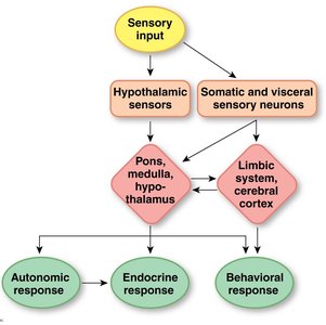
Autonomic Reflexes and Homeostasis
Autonomic reflexes are essential for maintaining homeostasis. The ANS works closely with the endocrine and behavioral systems to regulate physiological parameters such as blood pressure, heart rate, and digestion.
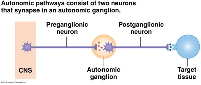
Organization of Autonomic Pathways
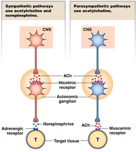
Autonomic pathways typically consist of two efferent neurons in series: a preganglionic neuron and a postganglionic neuron. Divergence is common, with one preganglionic neuron synapsing with multiple postganglionic neurons. The sympathetic and parasympathetic branches originate in different regions of the CNS and have distinct anatomical features.
Chemical Signaling in the Autonomic Nervous System
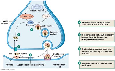
The ANS uses a variety of neurotransmitters and receptors. Most sympathetic postganglionic neurons release norepinephrine, while most parasympathetic postganglionic neurons release acetylcholine. Some neurons use other neurotransmitters such as substance P, ATP, or nitric oxide.
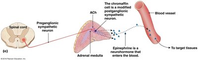
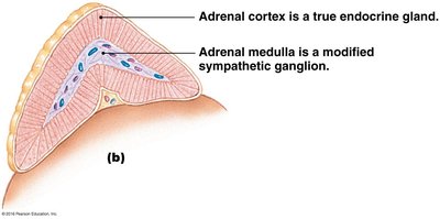
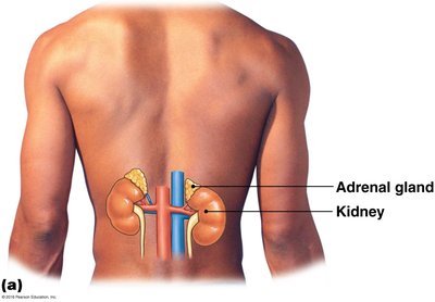
The Adrenal Medulla
The adrenal medulla is a specialized neuroendocrine structure associated with the sympathetic nervous system. It releases epinephrine (adrenaline) into the bloodstream, which acts on adrenergic receptors throughout the body.
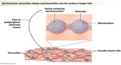
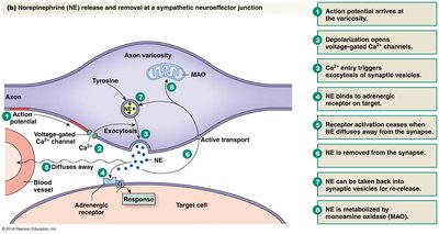
Neuroeffector Junctions and Neurotransmitter Release
Autonomic neurotransmitters are synthesized in the axon terminals and released at neuroeffector junctions. The primary neurotransmitters are acetylcholine (ACh) and norepinephrine (NE). Their release is calcium-mediated, and their action is terminated by reuptake or enzymatic breakdown.
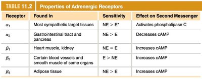
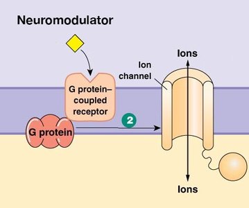
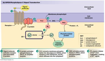
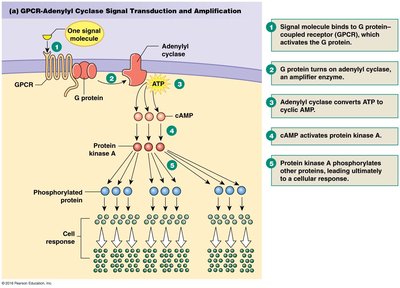
Autonomic Receptors and Signal Transduction
Autonomic receptors are primarily G-protein coupled receptors (metabotropic). Sympathetic adrenergic receptors are divided into alpha and beta subtypes, each with distinct second messenger effects. Parasympathetic muscarinic receptors (M1-M5) also have diverse effects depending on their location and signaling pathway.
Receptor | Found in | Sensitivity | Effect on Second Messenger |
|---|---|---|---|
α1 | Most sympathetic target tissues | NE > E | Activates phospholipase C |
α2 | Gastrointestinal tract and pancreas | NE > E | Decreases cAMP |
β1 | Heart muscle, kidney | NE = E | Increases cAMP |
β2 | Certain blood vessels and smooth muscle of some organs | E > NE | Increases cAMP |
β3 | Adipose tissue | NE > E | Increases cAMP |
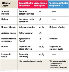
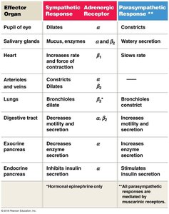
Summary Table: Autonomic Effects on Target Organs
Effector Organ | Sympathetic Response | Adrenergic Receptor | Parasympathetic Response |
|---|---|---|---|
Pupil of eye | Dilates | α | Constricts |
Salivary glands | Mucus, enzymes | α and β2 | Watery secretion |
Heart | Increases rate and force of contraction | β1 | Slows rate |
Arterioles and veins | Constriction/dilation | α, β2 | --- |
Lungs | Bronchioles dilate | β2 | Bronchioles constrict |
Digestive tract | Decreases motility and secretion | α, β2 | Increases motility and secretion |
Exocrine pancreas | Decreases enzyme secretion | α | Increases enzyme secretion |
Endocrine pancreas | Inhibits insulin secretion | α | Stimulates insulin secretion |
Adrenal medulla | Secretes catecholamines | --- | --- |
Kidney | Increases renin secretion | β1 | --- |
Urinary bladder | Urinary retention | α, β2 | Release of urine |
Adipose tissue | Fat breakdown | β3 | --- |
Male and female sex organs | Ejaculation (male) | α | Erection |
Uterus | Depends on stage of cycle | α, β2 | Depends on stage of cycle |
Lymphoid tissue | Generally inhibitory | α, β2 | --- |
Summary
Autonomic pathways consist of a preganglionic and postganglionic neuron in series, except for the adrenal medulla.
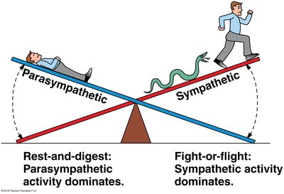
Sympathetic division is active during stress (fight-or-flight), while parasympathetic is active during rest-and-digest activities.
Neurotransmitter and receptor types determine the specific effects on target tissues.
