 Back
BackSkeletal and Muscle Systems: Structure, Function, and Organization
Study Guide - Smart Notes
 Tailored notes based on your materials, expanded with key definitions, examples, and context.
BONE TISSUE & STRUCTURE
Introduction to Bone Tissue
The skeletal system is composed of several tissue types, including bone (osseous tissue), cartilage, bone marrow, joint tissues, and periosteum. It serves multiple essential roles in the human body.
Support: Bones provide structural support, maintaining body posture.
Leverage: Bones act as levers for muscle action, enabling movement.
Protection: The skull, vertebral column, and rib cage protect vital organs.
Mineral Homeostasis: Bones store minerals such as calcium and phosphate, crucial for nerve conduction, muscle contraction, and blood clotting.
Blood Production: Red bone marrow produces red and white blood cells.
Energy Storage: Yellow bone marrow stores fat; brown marrow is present in children.
Bone Structure & Function
Bones are classified by shape (long, short, flat, irregular, sesamoid, sutural) and contain compact and spongy bone tissue. Long bones, such as those in the limbs, have distinct regions:
Epiphysis: Enlarged ends with spongy bone core and compact bone covering.
Diaphysis: Central shaft, mainly compact bone, surrounds the medullary cavity.
Metaphysis: Region between epiphysis and diaphysis; site of bone elongation at the epiphyseal plate.
Bone Marrow: Red marrow (hematopoietic) in spongy bone; yellow marrow (fat storage) in medullary cavity.
Periosteum: Dense fibrous membrane covering bone; contains osteogenic cells for growth and repair.
Articular Cartilage: Hyaline cartilage at joint surfaces, reducing friction and absorbing shock.
Bone Design
Thicker Bone at Stress Regions: Bone adapts to stress, being thicker where muscles attach and thinner elsewhere, optimizing strength and weight.
Matrix Composition: The matrix is a composite of hard calcium phosphate (hydroxyapatite) and flexible collagen fibers, providing both strength and flexibility.
Collagen Fiber Orientation: Collagen fibers in lamellae are arranged at varying angles, enhancing resistance to fracture.
Bone Cells
Osteoprogenitor Cells: Stem cells that differentiate into osteoblasts.
Osteoblasts: Bone-forming cells that secrete matrix and initiate calcification.
Osteocytes: Mature bone cells in lacunae, maintaining bone tissue.
Osteoclasts: Large, multinucleated cells that resorb bone matrix, remodeling bone.
Spongy (Cancellous) Bone Tissue
Spongy bone contains trabeculae (bony plates) with marrow-filled spaces, providing resistance to compression and reducing bone weight. It is prominent in short, flat, and irregular bones, and at the epiphyses of long bones.
Compact (Dense) Bone Tissue
Compact bone forms the outer layer of all bones and the shaft of long bones. It is organized into osteons (Haversian systems) with concentric lamellae around a central canal. Blood vessels and nerves travel through Haversian and Volkmann's canals. The endosteum lines internal bone surfaces and can generate new bone cells.
OSSIFICATION (BONE FORMATION)
Overview of Ossification
Bone formation occurs via two main processes:
Intramembranous Ossification: Forms flat bones within fibrous connective tissue (e.g., skull bones).
Endochondral Ossification: Forms most bones by replacing a cartilage model with bone (e.g., long bones).
Appositional Growth: Increases bone diameter by adding new bone to the surface.
Intramembranous Ossification
Begins in mesenchymal tissue; osteoprogenitor cells differentiate into osteoblasts, forming bone matrix around blood vessels.
Trabeculae form spongy bone; compact bone develops on surfaces.
Endochondral Ossification
Chondroblasts form a hyaline cartilage model, which grows and is gradually replaced by bone.
Primary ossification center forms in the diaphysis; secondary centers form in the epiphyses.
Epiphyseal plates allow for continued elongation until adulthood.
Bone Elongation at Epiphyseal Plate
Cartilage grows and is replaced by bone, lengthening the bone.
Osteoclasts digest calcified matrix; osteoblasts lay down new bone.
Appositional Growth
Osteoblasts in the periosteum add bone to the surface, increasing thickness.
Osteoclasts enlarge the medullary cavity as bone diameter increases.
FACTORS AFFECTING BONE GROWTH & DEVELOPMENT
Nutrition
Vitamins: A (osteoclast/osteoblast activity), B12 (osteoblast activity), C (collagen synthesis), D (calcium absorption).
Minerals: Calcium, phosphate, magnesium, manganese, boron.
Protein & Calories: Essential for collagen production and energy.
Hormones
Growth Hormone (GH): Stimulates bone elongation; abnormalities cause dwarfism or gigantism.
Thyroxine: Stimulates growth and ossification; excess or deficiency affects growth rate.
Calcitonin & Parathyroid Hormone: Regulate blood calcium by inhibiting or stimulating osteoclasts.
Sex Hormones: Stimulate bone growth, but prolonged exposure ossifies epiphyseal plates.
Insulin: Facilitates nutrient uptake for bone growth.
Exercise, Genetics, and Disease
Physical stress stimulates bone thickening; inactivity leads to atrophy.
Genetic factors and diseases can affect bone growth and health.
FRACTURES & BONE REPAIR
Types of Fractures
By Cause: Traumatic, pathologic, stress, compression.
By Skin Position: Closed (simple), open (compound).
By Fragment Number: Incomplete, complete, comminuted.
By Pattern: Hairline, greenstick, displaced, non-displaced, transverse, oblique, spiral, epiphyseal.
Fracture Repair
Hematoma formation, procallus (granulation tissue), internal/external callus, ossification, and remodeling.
Healing depends on blood supply, nutrients, and cell growth rate.
THE SKELETON
Axial vs. Appendicular Skeleton
Axial Skeleton (80 bones): Skull, vertebral column, sternum, ribs, ear bones, hyoid.
Appendicular Skeleton (126 bones): Pectoral girdle, upper limbs, pelvic girdle, lower limbs.
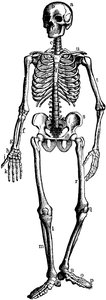
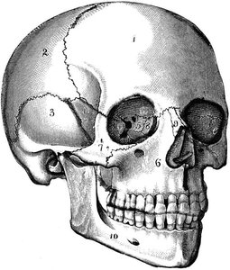
Skull
Cranium (8 bones): Frontal, parietal, temporal, occipital, sphenoid, ethmoid.
Face (14 bones): Nasal, maxillary, zygomatic, mandible, lacrimal, palatine, inferior nasal conchae, vomer.
Other Skeletal Regions
Auditory Ossicles & Hyoid: Malleus, incus, stapes, hyoid bone.
Spinal Column: Cervical (7), thoracic (12), lumbar (5), sacrum (1, fused), coccyx (1, fused).
Thoracic Cage: Sternum, ribs (true, false, floating).
Pectoral Girdle & Upper Appendages: Clavicle, scapula, humerus, radius, ulna, carpals, metacarpals, phalanges.
Pelvic Girdle & Lower Appendages: Ilium, ischium, pubis, femur, patella, tibia, fibula, tarsals, metatarsals, phalanges.
JOINTS (ARTICULATIONS)
Classification of Joints
By Structure: Fibrous (no cavity, e.g., sutures), cartilaginous (no cavity, e.g., pubic symphysis), synovial (joint cavity, e.g., knee).
By Function: Synarthrosis (immovable), amphiarthrosis (slightly movable), diarthrosis (freely movable).
Synovial Joints
Types: Gliding, hinge, pivot, condylar, saddle, ball & socket.
Structures: Articular capsule, synovial membrane, bursae, ligaments, articular disks (menisci), articular cartilage.
Movements: Flexion, extension, abduction, adduction, rotation, circumduction, supination, pronation, etc.
Joint Disorders
Arthritis (osteoarthritis, rheumatoid, gouty), bursitis, ankylosis, chondromalacia, dislocation, subluxation, sprain, strain, intervertebral disc disease.
MUSCLE TISSUE
Characteristics and Functions
Excitability, Contractility, Extensibility, Elasticity
Functions: Locomotion, posture, thermogenesis, glycogen storage, venous/lymph flow, breathing, speech, swallowing, defecation.
Organization of Skeletal Muscle
Muscle → Fascicles → Muscle fibers (cells) → Myofibrils → Sarcomeres (actin & myosin filaments).
Muscle fiber: Multinucleate, contains sarcolemma, T-tubules, sarcoplasmic reticulum, mitochondria.
Sarcomere: Z-lines, A-band (actin & myosin overlap), I-band (actin only), H-zone (myosin only), M-line.
Fascia & Connective Tissue Layers
Superficial fascia (hypodermis), deep fascia, epimysium (muscle), perimysium (fascicle), endomysium (fiber).
Tendons (muscle to bone), aponeuroses (muscle to muscle).
Neuronal Control & Membrane Potentials
Neuromuscular junction: Motor neuron releases acetylcholine, triggering muscle action potential.
Action potential: Rapid depolarization and repolarization of muscle cell membrane.
Muscle Cell Contraction
Stimulation by acetylcholine → Calcium release → Troponin/tropomyosin shift → Myosin-actin cross-bridge → Filament sliding → Sarcomere shortening.
Relaxation: Calcium reuptake, tropomyosin blocks actin, muscle returns to resting length.
Muscle Tension & Types of Contraction
Muscle twitch: Latent, contraction, relaxation phases.
Summation, tetanus, recruitment, muscle tone.
Isometric (tension without shortening), isotonic (shortening with movement: concentric/eccentric).
Muscle Energetics
ATP sources: Phosphagen system (creatine phosphate), anaerobic glycolysis, aerobic respiration.
Oxygen debt: Lactic acid buildup, post-exercise oxygen consumption.
Muscle fatigue: Depletion of ATP, glycogen, oxygen; lactic acid accumulation.
Skeletal Muscle Fiber Types
Slow (red, type I): Fatigue-resistant, high myoglobin, many mitochondria.
Fast-red (type IIa): Intermediate properties.
Fast (white, type IIb): Rapid, powerful, fatigue quickly, low myoglobin.
Smooth & Cardiac Muscle
Smooth muscle: Involuntary, non-striated, can regenerate, found in organs and vessels.
Cardiac muscle: Involuntary, striated, intercalated disks, cannot regenerate, found in heart.
MUSCLES & ACTIONS
Muscle Regions and Actions
Origin: Fixed attachment; Insertion: Movable attachment.
Belly: Largest part of muscle.
Agonist: Prime mover; Antagonist: Opposes agonist; Synergist: Assists movement.
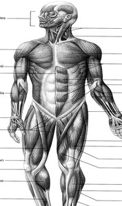
Major Muscle Groups
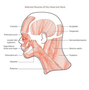
Head & Neck: Epicranius, orbicularis oculi/oris, zygomatic, masseter, temporalis, sternocleidomastoid.
Torso: Trapezius, latissimus dorsi, deltoid, pectoralis major, intercostals, diaphragm, abdominal muscles.
Arm: Biceps brachii, triceps brachii, brachioradialis.
Thigh: Sartorius, gracilis, gluteus maximus, biceps femoris, quadriceps femoris.
Lower Leg: Gastrocnemius, soleus, tibialis anterior.
Bone and Muscle Disorders
Bone: Osteoporosis, osteopenia, mastoiditis, osteomyelitis, osteogenesis imperfecta, lordosis, kyphosis, scoliosis.
Muscle: Atrophy, hypertrophy, multiple sclerosis, myasthenia gravis, myopathy, muscular dystrophy, poliomyelitis, ALS, fibrosis, fibrositis, fibromyalgia, paralysis from toxins (curare, pesticides, botulism).
Additional info: For more details on muscle contraction, see the sliding filament theory and the role of ATP in cross-bridge cycling. For bone repair, note the stages of callus formation and remodeling. For muscle fiber types, training can shift the proportion of fiber types in a muscle.
