 Back
BackSkeletal System: Structure, Organization, and Bone Types
Study Guide - Smart Notes
 Tailored notes based on your materials, expanded with key definitions, examples, and context.
Skeletal System Overview
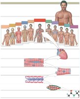
Levels of Organization in the Human Body
The human body is organized into hierarchical levels, each contributing to overall function and health. The skeletal system is one of several organ systems that work together to maintain life.
Organism Level: All organ systems function together for survival.
Organ System Level: Includes systems such as skeletal, muscular, nervous, cardiovascular, etc.
Organ Level: Organs are composed of multiple tissue types; for example, the heart contains muscle, connective, and nervous tissues.
Tissue Level: Groups of similar cells performing specific functions, such as cardiac muscle tissue.
Cellular Level: Cells are the basic units of life; heart muscle cells interlock to form tissue.
Chemical Level: Atoms and molecules form the structural and functional basis of cells.
Bone Tissue Types
Compact Bone vs. Spongy Bone
Bones are composed of two main types of tissue, each with distinct structural and functional properties.
Compact Bone (Dense Bone): Forms the outer surface of bones; provides strength where stress is applied in one direction.
Spongy Bone (Cancellous Bone): Located inside compact bone; lightweight and porous, reducing bone weight and housing marrow.
Structure of Bones
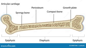
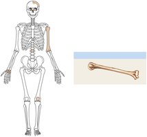
Long Bone Anatomy
Long bones, such as the femur, have a distinct anatomy that supports movement and growth.
Diaphysis: The shaft; composed mainly of compact bone.
Epiphysis: The ends; contain spongy bone and are covered with articular cartilage for joint movement.
Metaphysis: The region between diaphysis and epiphysis; contains the growth plate (epiphyseal plate in youth, epiphyseal line in adults).
Periosteum: Tough membrane covering bone; outer fibrous layer for muscle/ligament attachment, inner cellular layer produces osteoblasts.
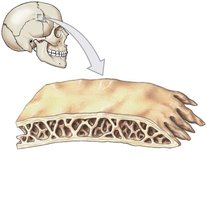
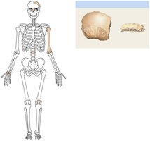
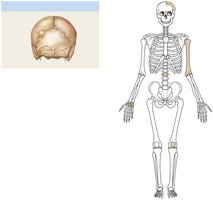
Flat Bone Anatomy
Flat bones, such as those in the skull, have a unique structure.
External Table: Outer layer of compact bone.
Diploë: Middle layer of spongy bone containing red marrow.
Internal Table: Inner layer of compact bone.
No marrow cavity: Red marrow in diploë produces blood cells.
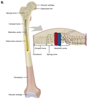
Internal Structure of Long Bones
Medullary (Marrow) Cavity: Hollow interior lined with spongy bone; contains yellow marrow (fat storage) and red marrow (blood cell production).
Endosteum: Membrane lining the cavity; contains osteoclasts for bone remodeling.
Bone Cells and Histology
Types of Bone Cells
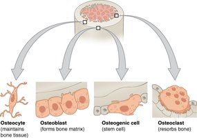
Bone tissue is maintained and remodeled by specialized cells.
Osteoblasts: Build bone matrix; active in growth and repair.
Osteocytes: Mature bone cells; maintain bone matrix.
Osteoclasts: Break down bone matrix for remodeling and mineral release.
Osteogenic Cells: Stem cells that differentiate into osteoblasts.
Compact Bone Histology
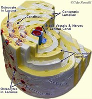
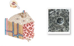
Compact bone is organized into structural units called osteons (Haversian systems).
Osteon: Column-like structure composed of concentric lamellae (rings of calcified matrix).
Lacunae: Small spaces containing osteocytes.
Canaliculi: Tiny channels for nutrient and waste exchange between osteocytes.
Central Canal: Contains blood vessels and nerves.
Perforating Canals: Connect central canals to periosteum and other osteons.
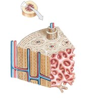

Spongy Bone Histology
Spongy bone lacks osteons and instead forms a lattice of trabeculae.
Trabeculae: Bony struts made of lamellae, intersected by canaliculi.
Spaces between trabeculae: Filled with red marrow, site of blood cell production.
Thin outer compact bone layer: Always seals spongy bone.
Organization of the Skeleton
Axial vs. Appendicular Skeleton
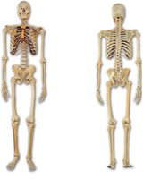
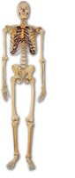
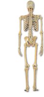
The adult skeleton consists of 206 bones, divided into two main parts.
Axial Skeleton (80 bones): Skull, vertebral column, sternum, ribs, hyoid bone; supports the central axis of the body.
Appendicular Skeleton (126 bones): Pectoral girdles, upper limbs, pelvic girdle, lower limbs; facilitates movement.
Classification of Bones by Shape
Bone Shapes and Examples
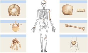
Bones are classified by their shapes, which relate to their functions.
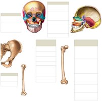
Long Bones: Greater in length than width; found in arms, forearms, thighs, legs (e.g., humerus, femur).
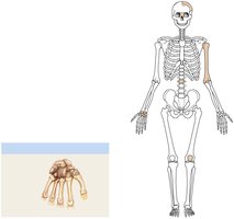
Short Bones: Nearly equal in length and width; found in wrists (carpals) and ankles (tarsals).
Flat Bones: Thin, plate-like; found in skull, ribs, sternum.
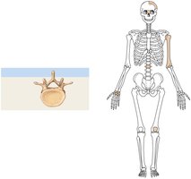
Irregular Bones: Complex shapes; found in vertebrae, some facial bones.
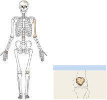
Sutural Bones: Small bones in skull sutures; number varies per person.
Sesamoid Bones: Form in tendons; example: patella.
Bone Markings
Categories of Bone Markings
Bone markings are anatomical features on bone surfaces, grouped into five categories.
General Structures: Body, head, neck.
Attachment Sites: Tuberosity, crest, spine; where tendons and ligaments attach.
Articulation Surfaces: Condyle, facet, trochlea; where bones meet.
Depressions: Fossa, sulcus; indentations for nerves or muscles.
Openings: Foramen, canal, fissure; passage for nerves and blood vessels.
Summary Table: Bone Types and Examples
Bone Type | Description | Example |
|---|---|---|
Long Bone | Longer than wide | Humerus, femur |
Short Bone | Nearly equal length and width | Carpals, tarsals |
Flat Bone | Thin, plate-like | Parietal bone, ribs |
Irregular Bone | Complex shape | Vertebra |
Sutural Bone | Small bones in skull sutures | Wormian bones |
Sesamoid Bone | Form in tendons | Patella |
Key Equations and Concepts
Bone Growth and Remodeling
Bone growth occurs at the epiphyseal plate, and remodeling is regulated by osteoblasts and osteoclasts.
Bone Matrix Composition: Mainly collagen fibers and hydroxyapatite (calcium phosphate).
Remodeling Equation:
Blood Cell Production
Red marrow in spongy bone is the site of hematopoiesis (blood cell formation).
Hematopoiesis Equation:
Conclusion
The skeletal system is a complex organ system essential for support, movement, protection, blood cell production, and mineral storage. Understanding bone structure, types, and markings is fundamental for anatomy and physiology studies.
