Back
BackSkin and Body Membranes: Structure, Function, and Clinical Relevance
Study Guide - Smart Notes
 Tailored notes based on your materials, expanded with key definitions, examples, and context.
Skin and Body Membranes
Overview of Body Membranes
Body membranes are essential structures that line body cavities, cover surfaces, and form protective barriers. They are classified into two main categories: epithelial membranes and connective tissue membranes.
Epithelial membranes include cutaneous, mucous, and serous membranes.
Connective tissue membranes primarily include synovial membranes.
Classification of Epithelial Tissues
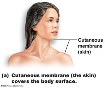
Cutaneous Membranes
The cutaneous membrane, commonly known as the skin, is the body's largest organ and serves as a protective covering.
Composed of two main layers: the epidermis (stratified squamous epithelium) and the dermis (dense connective tissue).
Functions as a barrier against mechanical, chemical, and microbial damage.
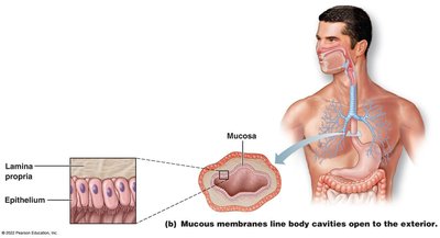
Mucous Membranes
Mucous membranes (mucosae) line all body cavities that open to the exterior, such as the respiratory, digestive, urinary, and reproductive tracts.
Composed of an epithelial layer overlying a connective tissue layer called the lamina propria.
Functions include secretion of mucus, absorption, and protection.
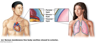
Serous Membranes
Serous membranes (serosae) line body cavities that are closed to the exterior, such as the thoracic and abdominal cavities.
Composed of simple squamous epithelium resting on areolar connective tissue.
Consist of two layers: parietal layer (lines cavity walls) and visceral layer (covers organs).
Layers are separated by serous fluid, reducing friction between organs.
Classification of Body Membranes: Connective Tissue
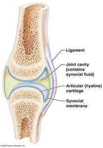
Synovial Membranes
Synovial membranes are specialized connective tissue membranes that line the fibrous capsules surrounding joints, bursae, and tendon sheaths.
Produce synovial fluid for lubrication and nourishment of joint cartilage.
Provide support, protection, and facilitate movement.
The Integumentary System
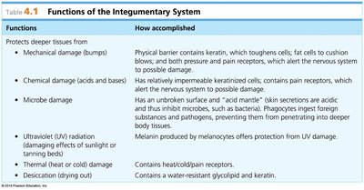
Functions of the Integumentary System
The integumentary system includes the skin, sweat and oil glands, hair, and nails. It serves as the body's primary barrier and plays multiple roles in protection, sensation, and regulation.
Functions | How accomplished |
|---|---|
Mechanical damage (bumps) | Physical barrier with keratin, fat cells, and pressure/pain receptors |
Chemical damage (acids and bases) | Keratinized cells, pain receptors |
Microbe damage | Unbroken surface, acid mantle, phagocytes |
Ultraviolet (UV) radiation | Melanin produced by melanocytes |
Thermal damage | Heat/cold/pain receptors |
Desiccation (drying out) | Water-resistant glycolipid and keratin |
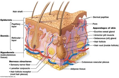
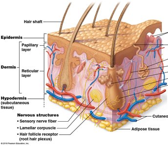
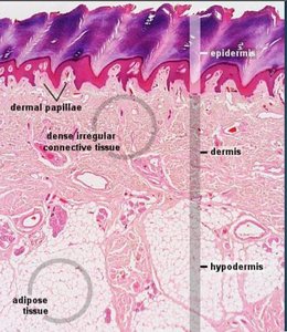
Structure of the Skin
Layers of the Skin
The skin is composed of three main layers: the epidermis, dermis, and hypodermis (subcutaneous tissue).
Epidermis: Outermost layer, primarily keratinocytes, provides waterproofing and protection.
Dermis: Middle layer, contains connective tissue, blood vessels, nerves, and appendages.
Hypodermis: Deepest layer, mainly adipose tissue, insulates and anchors skin.
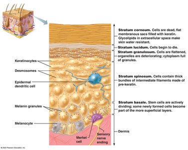
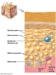
Epidermis
The epidermis consists of up to five layers, each with specialized cells and functions.
Keratinocytes: Produce keratin, a protein that strengthens and waterproofs the skin.
Melanocytes: Produce melanin, the pigment responsible for skin color.
Dendritic cells: Immune cells that detect pathogens.
Merkel cells: Sensory receptors for touch.
Dermis
The dermis is a strong, flexible connective tissue layer divided into two regions: the papillary layer and the reticular layer.
Papillary layer: Areolar connective tissue, forms dermal papillae (fingerprints), contains capillaries and sensory receptors.
Reticular layer: Dense irregular connective tissue, contains blood vessels, sweat and oil glands, collagen, and elastic fibers.
Blood vessels in the dermis help regulate temperature via vasodilation and vasoconstriction (negative feedback).
Homeostatic Imbalances of the Skin
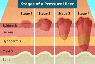
Pressure Ulcers (Decubitus Ulcers)
Pressure ulcers result from prolonged restriction of blood flow, leading to tissue death. They are common in immobile patients and progress through four stages.
Stage 1: Reddened skin, no open wounds.
Stage 2: Partial-thickness loss of dermis.
Stage 3: Full-thickness tissue loss, may expose fat.
Stage 4: Extensive destruction, may expose muscle or bone.
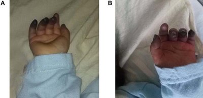
Cyanosis
Cyanosis is a bluish discoloration of the skin and mucous membranes due to low oxygen levels in the blood.
Commonly observed in lips, fingertips, and toes.
Indicates respiratory or circulatory issues.

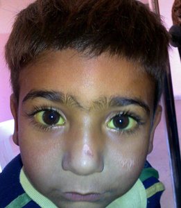
Jaundice
Jaundice is characterized by yellowing of the skin and eyes, caused by excess bile pigments in the blood, often due to liver dysfunction.
Indicates underlying hepatic or biliary disease.
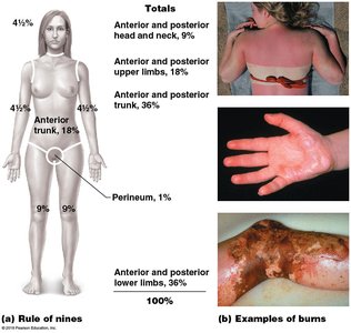
Burns
Burns are classified by depth and severity, ranging from first to fourth degree. Major concerns include fluid loss and infection.
First-degree: Affects only the epidermis.
Second-degree: Involves epidermis and part of dermis.
Third-degree: Destroys entire skin thickness.
Fourth-degree: Extends into deeper tissues.
Rule of Nines: Used to estimate body surface area affected by burns.
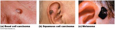
Skin Cancer
Skin cancer is the most common form of cancer. The ABCDE rule helps identify melanoma:
Asymmetry
Border irregularity
Color variation
Diameter > 6 mm
Evolving shape or size
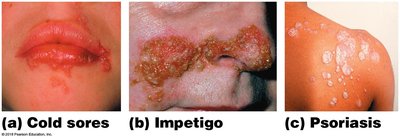
Infections and Allergies
Skin can be affected by infections (bacterial, viral) and allergic reactions. Common examples include cold sores, impetigo, and psoriasis.
Appendages of the Skin
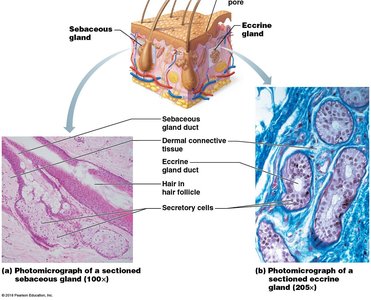
Cutaneous Glands
The skin contains two major types of glands: sebaceous (oil) glands and sweat (sudoriferous) glands.
Sebaceous glands: Produce sebum, which lubricates skin and hair, and has antibacterial properties. Overproduction can cause acne.
Sweat glands: Include eccrine (widely distributed, important for temperature regulation) and apocrine (found in armpits and genital areas, produce odor when broken down by bacteria).

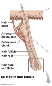
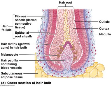
Hair and Hair Follicles
Hair is a flexible epithelial structure formed by the division of cells in the hair matrix. It consists of the medulla, cortex, and cuticle.
Cuticle: Outermost layer, provides strength and protection.
Hair follicle: Contains inner epithelial root sheath and outer fibrous sheath.
Hair papilla: Supplies blood to the growing hair.

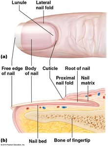
Nails
Nails are scale-like modifications of the epidermis. The nail matrix is responsible for growth, and the nail bed is the underlying epidermis. Nails appear pink due to the underlying blood supply, except for the lunule, which may appear blue in cases of cyanosis.