 Back
BackSkin and Membranes: Structure, Function, and Clinical Correlates
Study Guide - Smart Notes
 Tailored notes based on your materials, expanded with key definitions, examples, and context.
Skin & Membranes
Overview of Body Membranes
Body membranes are thin layers of tissue that cover surfaces, line body cavities, and form protective sheets around organs. They are classified based on their tissue composition and location in the body.
Epithelial membranes: Composed of epithelial tissue and an underlying layer of connective tissue.
Connective tissue membranes: Composed exclusively of various types of connective tissue.
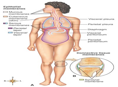
Types of Epithelial Membranes
Cutaneous membrane: The skin; primary organ of the integumentary system.
Serous membranes: Simple squamous epithelium on a connective tissue basement membrane. They line body cavities (parietal layer) and cover organs (visceral layer).
Mucous membranes: Line body surfaces that open directly to the exterior and produce mucus to keep membranes soft and moist.
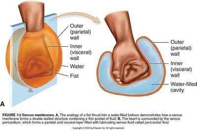
Examples of Serous Membranes
Pleura: Lines thoracic cavity and covers the lungs.
Peritoneum: Lines abdominal cavity and covers abdominal organs.
Diseases of Serous Membranes
Pleurisy (pleuritis): Inflammation of the pleura.
Peritonitis: Inflammation of the peritoneum.
Mucous Membranes
Contain both an epithelial layer and a fibrous connective tissue layer (lamina propria).
Examples: Lining of respiratory, digestive, urinary, and reproductive tracts.
Mucocutaneous junction: Transitional area where skin and mucous membranes meet.
Connective Tissue Membranes
Do not contain epithelial components.
Produce synovial fluid, a lubricant for joints and bursae.
Example: Synovial membranes in joint spaces and bursal sacs.
Skin Structure
Layers of the Skin
The skin (cutaneous membrane) is a sheetlike organ that acts as a barrier between the internal and external environment. It consists of two primary layers: the epidermis and the dermis.
Epidermis: Outermost, thinnest layer; composed of stratified squamous epithelium.
Dermis: Deeper, thicker layer; composed largely of connective tissue.
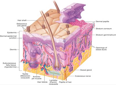
Epidermis
Stratum germinativum: Deepest layer; cells reproduce and move toward the surface.
Melanocytes: Produce melanin, the pigment responsible for skin color.
Keratinization: Cells fill with keratin, a tough, waterproof protein, as they move outward.
Stratum corneum: Outermost layer of keratin-filled cells.
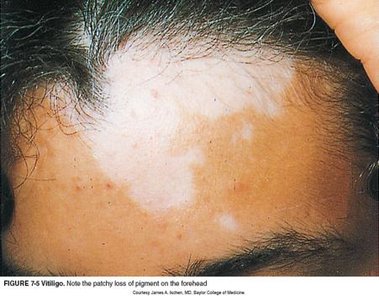
Skin Color Variations
Pink flush: Increased blood volume or oxygen.
Cyanosis: Bluish-gray color due to decreased blood oxygen.
Vitiligo: Patchy loss of melanocytes.
Albinism: Hereditary lack of melanin.
Freckles: Small, flat macules; normal pigment variation.
Dermal-Epidermal Junction
Specialized area of contact between epidermis and dermis; provides support.
Damage can cause blisters; widespread detachment is life-threatening due to infection risk.
Dermis
The dermis provides mechanical strength and houses various structures essential for skin function.
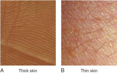
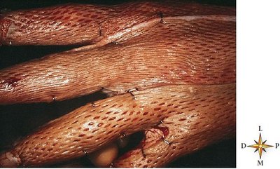
Contains parallel rows of dermal papillae (in thick skin, forms friction ridges).
Thick skin: Parallel friction ridges, no hair.
Thin skin: Irregular grooves, hair present.
Contains collagenous and elastic fibers; loss of elasticity with age causes wrinkles and striae (stretch marks).
Contains nerve endings, muscle fibers, hair follicles, sweat and sebaceous glands, and blood vessels.
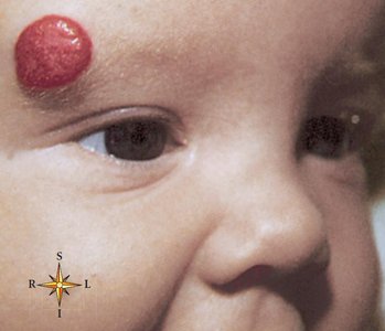
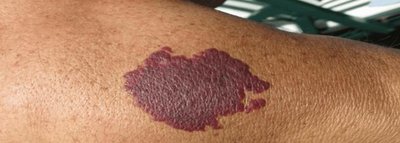
Birthmarks
Strawberry hemangioma: Mass of dilated dermal blood vessels.
Port-wine stain: Flat, red or purple mark present at birth.
Stork bite: Dilation of dermal capillaries at the nape of the neck in newborns.
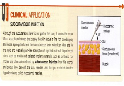
Subcutaneous Tissue (Hypodermis)
The subcutaneous tissue, or hypodermis, is not a true skin layer but connects the skin to underlying structures such as muscle and bone. It contains loose fibrous and adipose tissue and is a common site for subcutaneous injections.
Appendages of the Skin
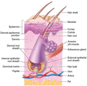
Hair
Lanugo: Soft hair of fetus and newborn.
Hair follicle: Epidermal tube-like structure required for hair growth.
Hair papilla: Cluster of cells where hair growth begins.
Hair root: Hidden in follicle; shaft: visible part.
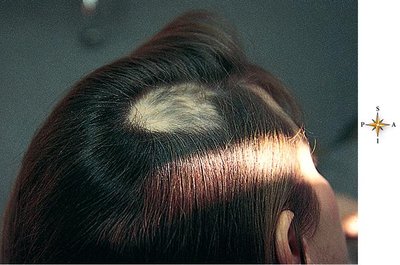
Alopecia: Hair loss.
Arrector pili: Smooth muscle causing "goose pimples" and hair to stand up.
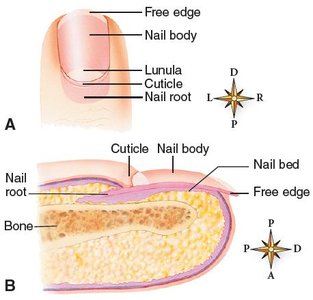
Nails
Produced by epidermal cells over terminal ends of fingers and toes.
Nail body: Visible part; root: hidden by cuticle.
Lunula: Crescent-shaped area near root.
Nail bed: May change color with blood flow changes.
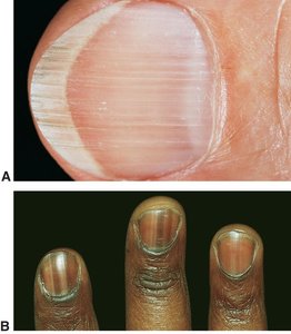
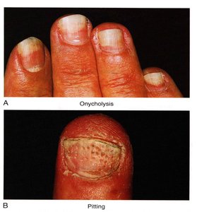
Variations in Nail Structure
Normal: Longitudinal ridges (light-skinned), pigmented bands (dark-skinned).
Abnormal: Onycholysis (separation from nail bed), pitting (common in psoriasis).
Receptors
Tactile (Meissner) corpuscle: Detects light touch.
Lamellar (Pacini) corpuscle: Detects pressure.
Skin Glands
Sweat (sudoriferous) glands: Eccrine (most numerous, regulate temperature, excrete waste) and apocrine (axilla/genitalia, thicker secretion, odor after bacterial breakdown).
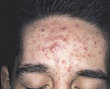
Sebaceous (oil) glands: Secrete sebum for lubrication; increased secretion during adolescence; can form blackheads and acne.
Functions of the Skin
Protection
First line of defense against microbes, UV rays, chemicals, cuts, and tears.
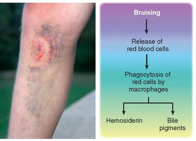
Bruising: Discoloration from blood vessel damage.
Skin grafts may be needed for severe damage.
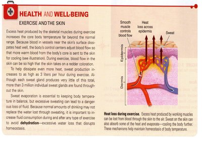
Temperature Regulation
Skin releases heat via sweat and blood flow regulation.
Can release up to 3000 calories of heat per day.
Sensation
Millions of nerve endings detect touch, pressure, pain, heat, and cold.
Excretion
Regulates sweat content; excretes uric acid, ammonia, urea, and excess substances.
Synthesis of Vitamin D
UV light stimulates production of vitamin D precursor, later converted in liver and kidneys.
Conditions of the Skin
Lesions
Elevated: Papule, plaque, vesicle, pustule, crust, wheal.
Flat: Macule.
Depressed: Excoriation, ulcer, fissure.
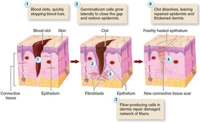
Skin Repair
Skin can repair itself after injury through clotting, cell migration, and tissue regeneration.
Burns
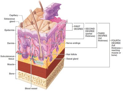
Classification and Severity
Severity depends on depth, total body surface area, and affected homeostatic mechanisms.
First-degree: Only epidermis affected.
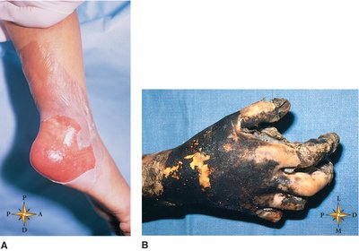
Second-degree: Deep epidermal and upper dermal layers affected.
Third-degree: Complete destruction of epidermis and dermis; may extend to muscle and bone (fourth-degree).
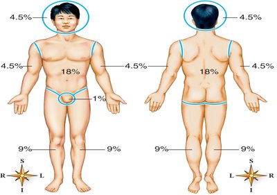
Rule of Nines
Used to estimate body surface area affected by burns; body divided into 11 areas of 9% each, plus 1% for genitals.
Skin Infections and Disorders
Common Infections
Impetigo: Contagious bacterial infection.
Tinea: Fungal infection.
Warts: Benign viral neoplasms.
Boils: Staphylococcal infection of hair follicles.
Scabies: Parasitic infection.
Vascular and Inflammatory Conditions
Decubitus ulcers: Bedsores from prolonged pressure.
Urticaria (hives): Red, itchy wheals from fluid loss.
Scleroderma: Autoimmune hardening of skin.
Psoriasis: Chronic, scaly plaques; genetic basis.
Eczema: Inflammatory condition with papules, vesicles, and crusts; symptom of underlying issue.
Skin Cancer
Squamous cell carcinoma: Slow-growing, may metastasize.
Basal cell carcinoma: Most common, rarely spreads.
Melanoma: Most serious, high mortality if untreated.
Kaposi sarcoma: Associated with immune deficiency, purple lesions.
ABCDE rule: For self-examination of moles (Asymmetry, Border, Color, Diameter, Evolving).
Review Questions
Which of these membranes is classified as a connective tissue membrane? Answer: Synovial
Identify the two main layers of the skin. Answer: Epidermis, dermis
The term _____ is used to describe a condition characterized by patchy looking areas of light skin resulting from the loss of epidermal melanocytes. Answer: Vitiligo
Hair growth begins from a small, cap-shaped cluster of cells called the: Answer: Hair papilla
Which of these would NOT be considered a function of the skin? Answer: Transportation
A burn that causes minor discomfort and some reddening of the skin is classified as a _____ burn. Answer: First-degree
Which skin disorder is characterized by silvery white, scale-like plaques that may remain fixed on the skin for months? Answer: Psoriasis
What is the most serious form of skin cancer? Answer: Melanoma
