 Back
BackStructure and Growth of Bones: An Overview for Anatomy & Physiology Students
Study Guide - Smart Notes
 Tailored notes based on your materials, expanded with key definitions, examples, and context.
Introduction to the Structure and Growth of Bones
Main Divisions and Functions of the Skeletal System
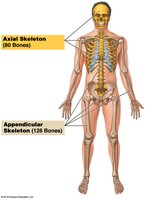
The human skeleton is divided into two main divisions: the axial skeleton and the appendicular skeleton. Each division plays a crucial role in supporting the body and facilitating movement.
Axial Skeleton: Composed of 80 bones, including the skull, thorax, and vertebral column. It forms the longitudinal axis of the body.
Appendicular Skeleton: Consists of 126 bones, including the limbs and girdles that attach them to the axial skeleton.
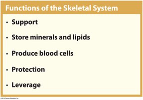
The skeletal system serves several vital functions:
Support: Provides structural support for the entire body.
Storage of Minerals and Lipids: Bones store calcium, phosphorus, and lipids in yellow marrow.
Blood Cell Production: Red marrow produces red blood cells, white blood cells, and platelets.
Protection: Protects internal organs (e.g., skull protects the brain, ribs protect the heart and lungs).
Leverage: Bones act as levers for muscle action, enabling movement.
Classification of Bones and Bone Markings
Bone Shapes and Examples
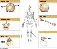
Bones are classified into six categories based on their shapes, each with specific examples and functions:
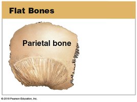
Flat Bones: Thin, parallel surfaces; protect soft tissues and provide muscle attachment (e.g., parietal bone of the skull).
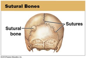
Sutural Bones: Small, irregular bones found between cranial bones (e.g., sutural bones in the skull).
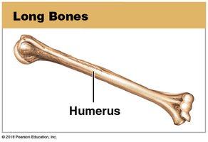
Long Bones: Long and slender; found in limbs (e.g., humerus).
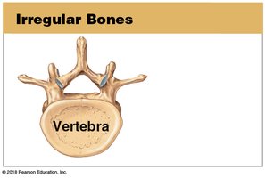
Irregular Bones: Complex shapes (e.g., vertebra).
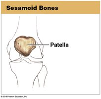
Sesamoid Bones: Small, round, and develop within tendons (e.g., patella).
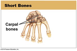
Short Bones: Small and boxy (e.g., carpal bones of the wrist).
Bone Markings (Surface Features)
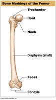
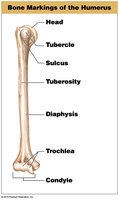
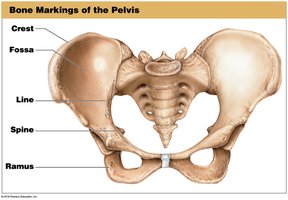
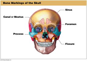
Bone markings are anatomical features on bones that serve as attachment points for muscles, tendons, and ligaments, or as passages for nerves and blood vessels. They are classified as elevations/projections or depressions/grooves/tunnels.
Elevations/Projections:
Process: Any projection or bump
Tubercle: Small, rounded projection
Tuberosity: Large, rough projection
Trochanter: Large, rough projection (femur only)
Condyle: Smooth, rounded articular process
Facet: Small, flat articular surface
Crest: Prominent ridge
Line: Low ridge
Spine: Pointed or narrow process
Ramus: Extension making an angle with the rest of the structure
Depressions/Grooves/Tunnels:
Canal or Meatus: Large passageway through a bone
Sinus: Chamber within a bone, usually filled with air
Foramen: Small, rounded passageway for blood vessels or nerves
Fissure: Elongated cleft or gap
Sulcus: Deep, narrow groove
Fossa: Shallow depression
Structure of a Typical Long Bone
Major Parts and Internal Structure
Long bones are specialized for transmitting forces and have a complex internal structure:
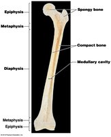
Epiphysis: Expanded area at each end, mostly spongy bone covered by compact bone; forms joints with other bones.
Metaphysis: Connects epiphysis to diaphysis; site of bone growth during development.
Diaphysis (Shaft): Long, tubular portion; contains the medullary (marrow) cavity.
Medullary Cavity: Central space containing red marrow (hematopoiesis) or yellow marrow (fat storage).
Articular Cartilage: Covers joint surfaces, reducing friction and absorbing shock.
Blood Supply and Innervation
Long bones require an extensive blood supply for growth and maintenance:
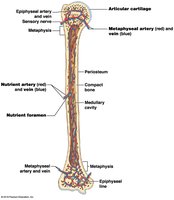
Nutrient Artery and Vein: Enter through the nutrient foramen to supply the diaphysis and medullary cavity.
Metaphyseal Arteries and Veins: Supply the metaphysis and connect to epiphyseal vessels.
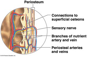
Periosteum: Contains smaller blood vessels, lymphatic vessels, and sensory nerves.
Bone Cells and Bone Matrix
Types of Bone Cells and Their Functions
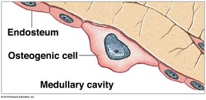
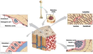
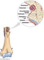
Osteogenic Cells: Stem cells that differentiate into osteoblasts; important for bone repair and found in the periosteum and endosteum.
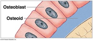
Osteoblasts: Produce new bone matrix (osteoid) and initiate calcification; become osteocytes when surrounded by matrix.
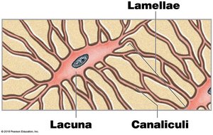
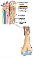
Osteocytes: Mature bone cells that maintain the bone matrix; reside in lacunae and communicate via canaliculi.
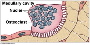
Osteoclasts: Large, multinucleated cells that resorb bone matrix, releasing stored minerals (osteolysis).
Bone Matrix Composition
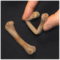
Collagen Fibers: Provide flexibility and tensile strength (~1/3 of bone weight).
Hydroxyapatite Crystals: Formed from calcium phosphate and calcium hydroxide; provide compressive strength (~2/3 of bone weight).
Other Components: Includes calcium carbonate and ions such as sodium, magnesium, and fluoride.
Compact Bone vs. Spongy Bone
Structure and Function
Compact Bone:
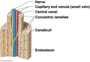
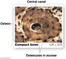
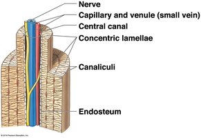
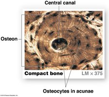
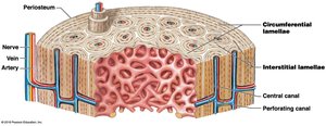
Functional unit is the osteon (Haversian system).
Concentric lamellae surround a central canal containing blood vessels and nerves.
Canaliculi connect osteocytes in lacunae to each other and to the central canal.
Strongest along its length; forms the outer layer of bones.
Spongy Bone:
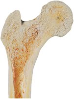
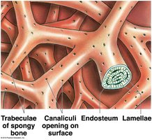
Composed of a network of trabeculae (struts and plates).
No osteons; lamellae form trabeculae with spaces for red marrow.
Nutrients reach osteocytes via canaliculi opening to the trabecular surface.
Found mainly in the epiphyses of long bones and inside flat bones.
Appositional Bone Growth
Mechanism and Effects
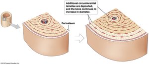
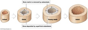
Appositional growth increases the diameter of bones by adding new layers of bone matrix under the periosteum. Osteogenic cells differentiate into osteoblasts, which deposit new circumferential lamellae. Osteoclasts resorb bone on the inner surface, enlarging the medullary cavity.
Periosteum and Endosteum
Periosteum: Outer fibrous layer and inner cellular layer; isolates bone, provides a route for blood vessels and nerves, and participates in growth and repair.
Endosteum: Incomplete cellular layer lining the medullary cavity; active in bone growth, repair, and remodeling.
Bone Formation: Ossification Processes
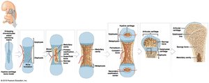
Endochondral Ossification
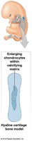
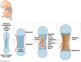
Most bones form by replacing a hyaline cartilage model with bone. This process involves several steps:
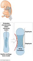
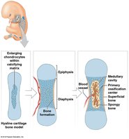
Cartilage model enlarges; chondrocytes die, leaving cavities.
Blood vessels grow around the cartilage; perichondrium cells become osteoblasts and form a superficial bone collar.
Blood vessels penetrate the cartilage; primary ossification center forms in the diaphysis.
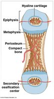
Medullary cavity forms as ossification spreads; secondary ossification centers develop in the epiphyses.
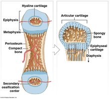
Epiphyses fill with spongy bone; articular cartilage and epiphyseal plate remain.
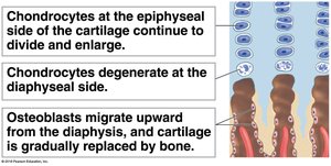
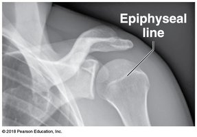
Bone grows in length at the epiphyseal plate until epiphyseal closure at adulthood.
Intramembranous Ossification
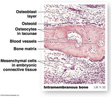
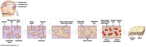
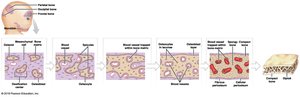
This process forms bone directly from mesenchymal (stem) cells, without a prior cartilage model. It is responsible for forming flat bones of the skull, mandible, clavicle, and some sesamoid bones.
Mesenchymal cells cluster and differentiate into osteoblasts, forming an ossification center.
Osteoblasts secrete osteoid, which becomes mineralized; trapped osteoblasts become osteocytes.
Bone grows outward in small struts (spicules); blood vessels invade and become trapped as spicules fuse.
Spongy bone forms; outer layers may be remodeled into compact bone.
Clinical Module: Abnormalities of Bone Growth
Disorders Affecting Bone Length
Pituitary Growth Failure: Inadequate growth hormone leads to reduced epiphyseal cartilage activity and short bones. Rare in the U.S. due to synthetic hormone therapy.
Achondroplasia: Slow growth of epiphyseal cartilage; results in short limbs but normal trunk size.
Marfan Syndrome: Excessive cartilage formation at epiphyseal plates; results in tall stature and long limbs; associated with cardiovascular issues.
Gigantism: Overproduction of growth hormone before puberty; causes excessive bone lengthening.
Acromegaly: Overproduction of growth hormone after epiphyseal plates close; bones thicken, especially in the face, jaw, and hands.
Congenital Talipes Equinovarus (Clubfoot): Inherited abnormality causing feet to turn medially and invert; treated with casts or supports.
Fibrodysplasia Ossificans Progressiva (FOP): Genetic disorder causing bone to form in muscles and other soft tissues (heterotopic ossification).
Physiology of Bones: Mineral Storage and Homeostasis
Mineral Storage and Calcium Homeostasis
Minerals in Bone: Bones store calcium, phosphate, and other ions essential for physiological processes.
Calcium: Most abundant mineral in the body; ~99% stored in bones. Essential for muscle contraction, nerve function, and blood clotting.
Organs Involved in Calcium Homeostasis:
Intestines: Absorb calcium and phosphate under hormonal control.
Bones: Osteoclasts release calcium; osteoblasts deposit calcium.
Kidneys: Regulate calcium and phosphate loss in urine.
Hormonal Regulation of Calcium
Parathyroid Hormone (PTH): Increases blood calcium by stimulating osteoclasts, increasing intestinal absorption (via calcitriol), and reducing urinary loss.
Calcitriol: Active form of vitamin D; enhances calcium absorption in the intestines.
Calcitonin: Lowers blood calcium by inhibiting osteoclasts, decreasing intestinal absorption, and increasing urinary excretion.
Clinical Module: Bone Fractures and Repair
Types of Fractures
Closed (Simple) Fracture: Bone does not break the skin.
Open (Compound) Fracture: Bone projects through the skin; higher risk of infection and bleeding.
Transverse Fracture: Break across the long axis.
Spiral Fracture: Produced by twisting stresses.
Displaced/Nondisplaced Fractures: Displaced fractures produce abnormal bone arrangements; nondisplaced retain normal alignment.
Compression Fracture: Occurs in vertebrae under extreme stress.
Greenstick Fracture: One side of the shaft breaks, the other bends; common in children.
Comminuted Fracture: Bone shatters into fragments.
Epiphyseal Fracture: Occurs at the epiphyseal plate; may affect bone growth if not properly managed.
Pott’s (Bimalleolar) Fracture: Affects both malleoli of the ankle.
Colles Fracture: Break in the distal radius.
Steps in Fracture Repair
Fracture Hematoma Formation: Blood clot forms at the fracture site.
Callus Formation: Internal callus (spongy bone) unites inner edges; external callus (cartilage and bone) stabilizes outer edges.
Spongy Bone Formation: Cartilage of external callus is replaced by spongy bone; dead bone is removed.
Compact Bone Formation: Spongy bone is remodeled into compact bone; bone regains normal shape and structure.
Example: A transverse fracture of the femur will heal through these four steps, provided the blood supply and periosteum/endosteum are intact.
Additional info: The process of bone repair can take weeks to months, depending on the severity of the fracture and the individual's age and health status.
