 Back
BackStudy Guide: Bones and Bone Structure in Anatomy & Physiology
Study Guide - Smart Notes
 Tailored notes based on your materials, expanded with key definitions, examples, and context.
Bones and Bone Structure
Functions of Bone
Bones are specialized connective tissues that serve multiple essential functions in the human body. They provide structural support, facilitate movement, protect internal organs, store minerals, and participate in blood cell production.
Support: Bones form the framework that supports the body and anchors soft tissues.
Protection: Bones protect vital organs, such as the brain (skull), heart and lungs (rib cage), and spinal cord (vertebrae).
Movement: Bones act as levers for muscles, enabling movement at joints.
Mineral Storage: Bones store minerals, primarily calcium and phosphate, which are released into the bloodstream as needed.
Blood Cell Production: The bone marrow produces red blood cells, white blood cells, and platelets (hematopoiesis).
Energy Storage: Yellow bone marrow stores lipids as an energy reserve.
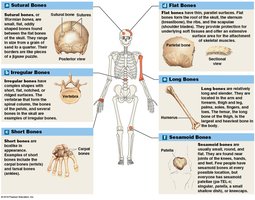
Classification of Bones
Bones are classified based on their shape and structure, which relates to their function and location in the body.
Sutural Bones: Small, irregular bones found between the flat bones of the skull.
Irregular Bones: Complex shapes, such as vertebrae and pelvic bones.
Short Bones: Small and boxy, like the bones of the wrist (carpals) and ankle (tarsals).
Flat Bones: Thin, parallel surfaces; examples include the skull, ribs, and scapulae.
Long Bones: Longer than they are wide; examples include the femur, humerus, and tibia.
Sesamoid Bones: Small, round bones embedded within tendons, such as the patella.
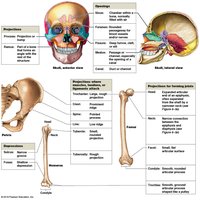
Bone Markings and Surface Features
Bones display various markings and features that serve as attachment points for muscles, tendons, and ligaments, and allow passage for nerves and blood vessels.
Projections: Sites for muscle and ligament attachment (e.g., tuberosity, crest, trochanter).
Openings: Allow passage of blood vessels and nerves (e.g., foramen, canal).
Depressions: Areas that accommodate other structures (e.g., fossa, groove).
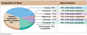
Composition of Bone
Bones are composed of both organic and inorganic components, which provide strength, flexibility, and mineral storage.
Organic Compounds: Mostly collagen fibers, providing flexibility and tensile strength.
Inorganic Compounds: Mainly hydroxyapatite (calcium phosphate crystals), providing hardness and resistance to compression.
Mineral Content: Bones contain most of the body's calcium, phosphate, and significant amounts of other minerals.
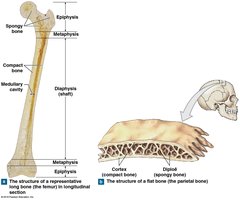
Structure of Long and Flat Bones
Long bones and flat bones have distinct structural features that reflect their functions.
Long Bones: Consist of a diaphysis (shaft), epiphyses (ends), metaphysis (growth region), compact bone, spongy bone, and medullary cavity.
Flat Bones: Composed of two layers of compact bone with a spongy bone (diploë) in between.
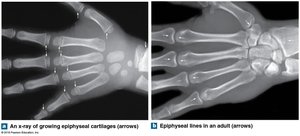
Epiphyseal Cartilage and Growth
Epiphyseal cartilage (growth plate) is responsible for longitudinal bone growth during childhood and adolescence. In adults, the cartilage is replaced by bone, forming the epiphyseal line.
Epiphyseal Cartilage: Visible in growing bones; allows for lengthening.
Epiphyseal Line: Remnant in adults after growth has ceased.
Spongy Bone and Osteoporosis
Spongy bone (trabecular bone) is characterized by a porous, lattice-like structure. Osteoporosis is a condition where bone density decreases, increasing fracture risk.
Normal Spongy Bone: Dense, interconnected trabeculae.
Osteoporotic Bone: Thinner, less connected trabeculae, leading to fragility.

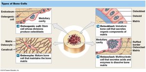
Types of Bone Cells
Bone tissue contains several cell types, each with specific functions in bone formation, maintenance, and remodeling.
Osteogenic Cells: Stem cells that differentiate into osteoblasts.
Osteoblasts: Cells that synthesize new bone matrix.
Osteocytes: Mature bone cells that maintain bone tissue.
Osteoclasts: Multinucleated cells that break down bone matrix.
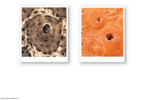
Microscopic Structure of Compact Bone
Compact bone is organized into osteons (Haversian systems), which are cylindrical structures containing concentric lamellae, central canals, lacunae, and canaliculi.
Osteon: Functional unit of compact bone.
Lamellae: Layers of bone matrix.
Central Canal: Contains blood vessels and nerves.
Lacunae: Small spaces housing osteocytes.
Canaliculi: Tiny channels connecting lacunae.
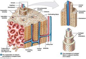
Organization of Compact Bone
Compact bone is organized to optimize strength and facilitate nutrient delivery. Collagen fibers in adjacent lamellae are oriented in different directions, increasing resistance to torsion.
Perforating Canals: Connect central canals and provide pathways for blood vessels.
Collagen Fiber Orientation: Alternating directions in lamellae enhance strength.
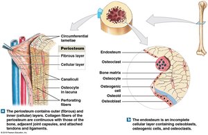
Periosteum and Endosteum
The periosteum and endosteum are connective tissue membranes that cover bone surfaces and participate in bone growth and repair.
Periosteum: Covers outer surface of bone; consists of fibrous and cellular layers.
Endosteum: Lines internal bone surfaces; contains osteogenic cells, osteoblasts, and osteoclasts.
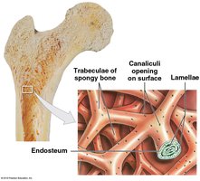
Spongy Bone Structure
Spongy bone consists of trabeculae, which are oriented along lines of stress. Canaliculi open onto the surface, allowing osteocytes to exchange nutrients.
Trabeculae: Lattice-like network providing structural support.
Endosteum: Lines trabeculae and participates in bone remodeling.
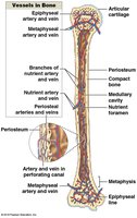
Vascular Supply of Bone
Bones are highly vascularized, with arteries and veins supplying nutrients and removing waste. Blood vessels enter through nutrient foramina and branch throughout the bone.
Periosteal Vessels: Supply outer compact bone.
Nutrient Artery and Vein: Enter through the diaphysis.
Metaphyseal and Epiphyseal Vessels: Supply the ends of bones.
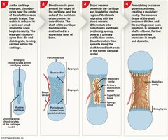
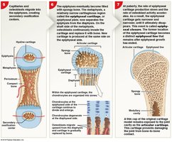
Bone Growth and Development
Bone growth occurs through endochondral ossification, where cartilage is replaced by bone. This process involves several stages, including cartilage model formation, vascular invasion, and bone remodeling.
Endochondral Ossification: Most bones form by replacing hyaline cartilage with bone tissue.
Growth Plate: Site of longitudinal growth in children.
Remodeling: Bone is continuously renewed throughout life.
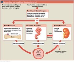
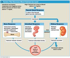
Regulation of Blood Calcium Levels
Blood calcium levels are tightly regulated by hormones, primarily parathyroid hormone (PTH) and calcitonin. These hormones control calcium release from bone, absorption in the intestine, and excretion by the kidneys.
Low Calcium: PTH increases calcium release from bone, absorption in intestine, and retention in kidneys.
High Calcium: Calcitonin decreases calcium release from bone, absorption in intestine, and increases excretion in kidneys.
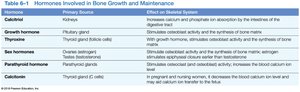
Hormones Involved in Bone Growth and Maintenance
Several hormones influence bone growth, remodeling, and maintenance. These include calcitriol, growth hormone, thyroxine, sex hormones, parathyroid hormone, and calcitonin.
Hormone | Primary Source | Effect on Skeletal System |
|---|---|---|
Calcitriol | Kidneys | Increases calcium and phosphate absorption from digestive tract |
Growth hormone | Pituitary gland | Stimulates osteoblast activity and bone synthesis |
Thyroxine | Thyroid gland | With growth hormone, stimulates osteoblast activity and bone synthesis |
Sex hormones | Ovaries/testes | Stimulate osteoblast activity and synthesis of bone matrix; estrogen stimulates closure of epiphyseal plates |
Parathyroid hormone | Parathyroid glands | Stimulates osteoclast activity; increases blood calcium level |
Calcitonin | Thyroid gland (C cells) | In pregnant/lactating women, decreases blood calcium level and may limit calcium transfer to fetus |
Key Terms and Concepts
Osteon (Haversian system): The structural unit of compact bone.
Lamellae: Concentric rings of bone matrix within an osteon.
Lacunae: Small spaces containing osteocytes.
Canaliculi: Microscopic channels connecting lacunae.
Trabeculae: Lattice-like structures in spongy bone.
Periosteum: Membrane covering the outer surface of bone.
Endosteum: Membrane lining internal bone surfaces.
Epiphyseal Plate: Growth plate in long bones.
Hematopoiesis: Blood cell formation in bone marrow.
Equations and Formulas
Bone mineral density and calcium homeostasis are important concepts in bone physiology.
Calcium Homeostasis Equation:
Additional info: Academic context was added to clarify bone functions, cell types, and regulatory mechanisms, as well as to expand on the brief points and provide self-contained explanations suitable for exam preparation.
