 Back
BackStudy Guide: Histology, Integumentary System, and Bone Structure (Ch. 4–6, ANP College Level)
Study Guide - Smart Notes
 Tailored notes based on your materials, expanded with key definitions, examples, and context.
Chapter 4: The Tissue Level of Organization (Histology)
Epithelial Tissues
Epithelial tissues cover body surfaces, line cavities, and form glands. They are specialized for protection, absorption, secretion, and sensation.
Key Features: High cellularity, polarity (apical/basal), attachment to basement membrane, avascularity, rapid regeneration.
Specializations: Cilia, microvilli, secretory cells, germinative (basal) layer.
Classification of Epithelia
By Layers: Simple (one layer), Stratified (multiple layers)
By Shape: Squamous (flat), Cuboidal (cube), Columnar (tall)
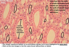
Simple Squamous Epithelium
Structure: Single layer of flat cells
Locations: Alveoli, blood vessels (endothelium), serous membranes (mesothelium), kidney tubules
Functions: Diffusion, filtration, reduces friction
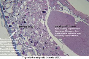
Simple Cuboidal Epithelium
Structure: Single layer of cube-shaped cells
Locations: Glands, ducts, kidney tubules, thyroid gland
Functions: Secretion, absorption, limited protection
Simple Columnar Epithelium
Structure: Single layer of tall cells, often with microvilli or goblet cells
Locations: Stomach, intestines, gallbladder, uterine tubes
Functions: Protection, secretion, absorption
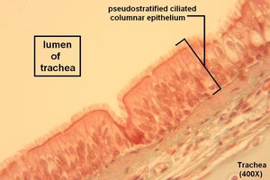
Pseudostratified Columnar Epithelium
Structure: Appears layered but all cells touch basement membrane; often ciliated with goblet cells
Locations: Trachea, bronchi, nasal cavity, male reproductive tract
Functions: Protection, secretion, movement of mucus by cilia
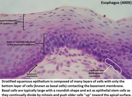
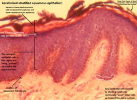
Stratified Squamous Epithelium
Structure: Multiple layers; surface cells are flat
Locations: Skin (keratinized), mouth, esophagus, anus, vagina (non-keratinized)
Functions: Protection against abrasion, pathogens, chemicals
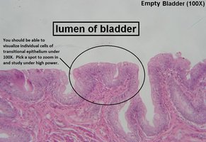
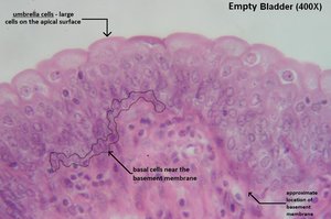
Transitional Epithelium
Structure: Multiple layers; cells change shape (dome-shaped when relaxed, flat when stretched)
Locations: Urinary bladder, ureters, renal pelvis
Functions: Permits expansion and recoil after stretching
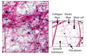
Connective Tissues (CT)
Connective tissues support, bind, and protect organs. They have specialized cells, protein fibers, and ground substance (matrix).
Cell Types: Fibroblasts (make fibers), adipocytes (store fat), macrophages (phagocytes)
Fiber Types: Collagen (strong), elastic (stretchy), reticular (network)
Areolar (Loose) Connective Tissue
Structure: Loose arrangement of fibers, abundant ground substance
Locations: Under epithelia, around organs
Functions: Cushions organs, provides support, allows movement
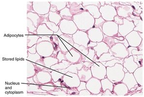
Adipose Tissue
Structure: Large, closely packed adipocytes (fat cells)
Locations: Under skin, around eyes/kidneys, within abdomen
Functions: Energy storage, insulation, cushioning
Dense Regular Connective Tissue
Structure: Parallel collagen fibers, few cells
Locations: Tendons, ligaments
Functions: Strong attachment, resists pulling forces in one direction
Dense Irregular Connective Tissue
Structure: Collagen fibers in multiple directions
Locations: Dermis, organ capsules
Functions: Withstands tension from many directions
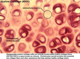
Cartilage (Supportive CT)
General Features: Chondrocytes in lacunae, avascular, firm matrix
Types: Hyaline (most common), elastic, fibrocartilage
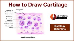
Hyaline Cartilage
Structure: Glassy matrix, chondrocytes in lacunae
Locations: Ends of long bones, nose, trachea, larynx, costal cartilage
Functions: Support, reduces friction, absorbs shock
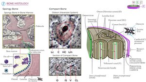
Bone (Osseous Tissue)
Structure: Osteocytes in lacunae, hard mineralized matrix, organized in osteons (compact bone)
Functions: Support, protection, movement, mineral storage, blood cell formation

Membranes
Mucous Membranes: Line passages open to exterior; secrete mucus
Serous Membranes: Line closed cavities; secrete serous fluid
Cutaneous Membrane: Skin
Synovial Membranes: Line joint cavities; secrete synovial fluid
Muscle Tissue
Skeletal Muscle: Voluntary, striated, multinucleate
Cardiac Muscle: Involuntary, striated, branched, intercalated discs
Smooth Muscle: Involuntary, non-striated, spindle-shaped cells
Nervous Tissue
Neurons: Conduct electrical impulses; have cell body, dendrites, axon
Neuroglia: Support, protect, and nourish neurons
Tissue Injury and Repair
Inflammation: Response to injury; involves swelling, redness, heat, pain
Regeneration: Replacement of damaged tissue; varies by tissue type
Chapter 5: The Integumentary System (Skin)
Overview and Functions
The integumentary system includes the skin and its accessory structures (hair, nails, glands). It protects the body, regulates temperature, synthesizes vitamin D, and provides sensation.
Protection: Physical, chemical, and biological barrier
Excretion: Sweat removes wastes
Storage: Fat, water, vitamins
Sensation: Touch, pain, temperature
Vitamin D Synthesis: UV light converts precursor to vitamin D3
Thermoregulation: Sweat and blood flow control heat loss
Skin Structure
Epidermis: Keratinized stratified squamous epithelium; main cell = keratinocyte
Dermis: Connective tissue (papillary = areolar; reticular = dense irregular)
Hypodermis: Subcutaneous layer; adipose + areolar tissue
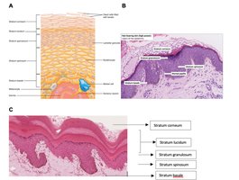
Epidermal Layers (Deep to Superficial)
Stratum basale (germinativum): Stem cells, melanocytes, Merkel cells
Stratum spinosum: Keratinocytes, Langerhans cells
Stratum granulosum: Keratin production, cells begin to die
Stratum lucidum: Only in thick skin (palms, soles)
Stratum corneum: Dead, keratinized cells; barrier layer
Other Epidermal Cells
Melanocytes: Produce melanin (UV protection)
Dendritic (Langerhans) cells: Immune defense
Tactile (Merkel) cells: Sensory receptors
Skin Color Factors
Melanin: Brown-black pigment from melanocytes
Carotene: Yellow-orange pigment
Hemoglobin: Red pigment in blood
Accessory Structures
Hair: Protection, insulation, sensation
Sebaceous glands: Secrete sebum (oil)
Sudoriferous glands: Eccrine (watery sweat), apocrine (thicker, odor)
Nails: Protect digits, aid grasping
Burns (by Depth)
1st degree: Epidermis only; red, no scar
2nd degree: Epidermis + part of dermis; blisters, may scar
3rd degree: Full thickness; white/charred, scars, infection risk
Skin Cancer
Basal cell carcinoma: Most common, least dangerous
Squamous cell carcinoma: Can metastasize
Malignant melanoma: Most deadly; from melanocytes
Aging Effects
Thinner skin, less elasticity, slower healing, gray hair, less sweat/sebum
Chapter 6: Osseous Tissue & Bone Structure
Functions of Bone
Support, protection, movement, mineral storage, blood cell formation (hematopoiesis)
Classification by Shape
Long, short, flat, irregular, sesamoid, sutural
Structure of Long Bones
Diaphysis: Shaft; compact bone, medullary cavity (yellow marrow)
Epiphysis: Ends; spongy bone (red marrow), articular cartilage
Metaphysis: Growth zone (epiphyseal plate in children)
Bone Matrix
Inorganic: 2/3 calcium phosphate (hydroxyapatite) – hardness
Organic: 1/3 collagen fibers – flexibility
Bone Cells
Osteoprogenitor: Stem cells
Osteoblasts: Build bone matrix
Osteocytes: Maintain matrix, in lacunae
Osteoclasts: Break down bone (resorption)
Compact Bone Structure
Organized in osteons (Haversian systems): central canal, concentric lamellae, lacunae, canaliculi, perforating canals
Periosteum: Outer covering; Endosteum: Lines medullary cavity
Bone Development & Growth
Osteogenesis: Bone formation
Intramembranous ossification: Flat bones (skull)
Endochondral ossification: Most bones; cartilage model replaced by bone
Epiphyseal plate: Growth in length; closes at adulthood (epiphyseal line)
Fracture Repair
Hematoma (blood clot)
Fibrocartilaginous (soft) callus
Bony (hard) callus
Remodeling
Bone Homeostasis
Balance between osteoblast and osteoclast activity
Regulated by hormones (PTH, calcitonin, calcitriol), nutrition, exercise
Calcium Balance
PTH: Raises blood Ca2+
Calcitonin: Lowers blood Ca2+
Calcitriol: Increases Ca2+ absorption in intestines
Aging and Bone Disorders
Osteopenia: Bone thinning with age
Osteoporosis: Severe bone loss; increased fracture risk
Quick Exam Tips
Memorize tissue locations and functions
Know skin layers and color factors
Understand bone matrix and cell types
Practice identifying tissues from images
