 Back
BackLecture - Ch 12 Nervous System Fundamentals
Study Guide - Smart Notes
 Tailored notes based on your materials, expanded with key definitions, examples, and context.
The Nervous System
Overview and Functions
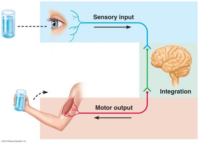
The nervous system is the master controlling and communicating system of the body, responsible for rapid and specific responses to internal and external stimuli. It operates through electrical and chemical signals, enabling immediate reactions.
Sensory Input: Information is gathered by sensory receptors about changes inside and outside the body.
Integration: The brain processes and interprets sensory input.
Motor Output: Activation of effector organs (muscles and glands) produces a response.
Divisions of the Nervous System
The nervous system is divided into two principal parts:
Central Nervous System (CNS): Consists of the brain and spinal cord. It is the integration and control center, interpreting sensory input and dictating motor output.
Peripheral Nervous System (PNS): Composed mainly of nerves extending from the CNS. It has two functional divisions:
Sensory (afferent) division: Somatic sensory fibers convey impulses from skin, skeletal muscles, and joints; visceral sensory fibers convey impulses from visceral organs.
Motor (efferent) division: Transmits impulses from CNS to effector organs. Includes the somatic nervous system (voluntary control of skeletal muscles) and the autonomic nervous system (involuntary control of smooth muscle, cardiac muscle, and glands; subdivided into sympathetic and parasympathetic divisions).
Nervous Tissue Histology
Cell Types
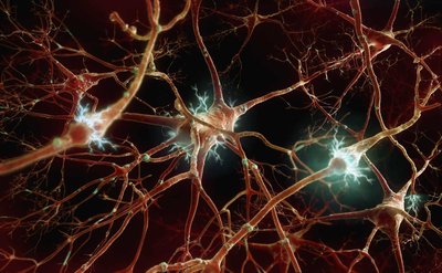
Nervous tissue consists of two principal cell types:
Neuroglia (glial cells): Support, protect, and insulate neurons. Types include astrocytes, microglial cells, ependymal cells, and oligodendrocytes in the CNS; satellite cells and Schwann cells in the PNS.
Neurons: Excitable cells that transmit electrical signals. They are large, highly specialized, and have extreme longevity, are mostly amitotic, and require continuous oxygen and glucose supply.
Neuroglia Functions
Astrocytes: Support neurons, regulate exchanges between capillaries and neurons, guide neuron migration, and control the chemical environment.
Microglial Cells: Monitor neuron health and can transform to phagocytize microorganisms and debris.
Ependymal Cells: Line CNS cavities, circulate cerebrospinal fluid (CSF).
Oligodendrocytes: Form myelin sheaths in CNS.
Satellite Cells: Surround neuron cell bodies in PNS, similar to astrocytes.
Schwann Cells: Form myelin sheaths in PNS, vital for regeneration of damaged nerve fibers.

Neurons: Structure and Function
Neuron Anatomy
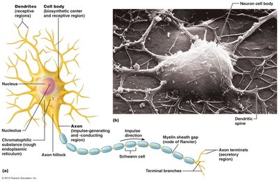
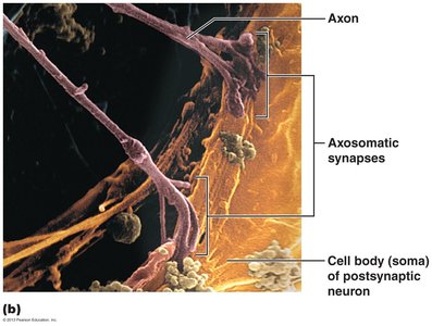
Neurons are the structural units of the nervous system, consisting of a cell body (soma) and processes (dendrites and axons).
Cell Body: Biosynthetic center, contains nucleus and organelles, receptive region for input.
Dendrites: Short, branched processes that receive signals and convey them toward the cell body as graded potentials.
Axon: Long process that conducts impulses away from the cell body, ending in axon terminals that release neurotransmitters.
Myelination
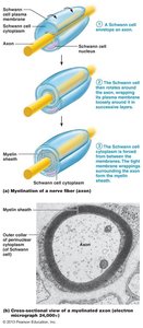
Myelin Sheath: Whitish, protein-lipid covering that insulates axons and increases impulse speed.
Schwann Cells: Form myelin in PNS by wrapping around axons.
Myelin Sheath Gaps (Nodes of Ranvier): Gaps between Schwann cells where axon collaterals can emerge.
Oligodendrocytes: Form myelin in CNS, can wrap multiple axons.
Classification of Neurons
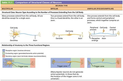
Structural Classification
Neurons are classified by the number of processes extending from the cell body:
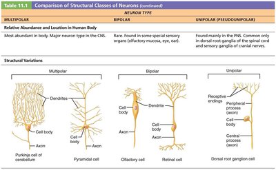
Multipolar: Three or more processes (one axon, others dendrites); most common in CNS.
Bipolar: Two processes (one axon, one dendrite); rare, found in retina and olfactory mucosa.
Unipolar (pseudounipolar): One T-like process; found mainly in PNS.
Neuron Type | Processes | Location |
|---|---|---|
Multipolar | Many dendrites, one axon | CNS |
Bipolar | One dendrite, one axon | Special sensory organs |
Unipolar | One T-like process | PNS |
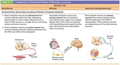
Functional Classification
Neurons are also classified by the direction of impulse conduction:
Sensory (afferent): Transmit impulses toward CNS; mostly unipolar.
Motor (efferent): Carry impulses from CNS to effectors; multipolar.
Interneurons: Shuttle signals within CNS; multipolar, most abundant.
Neurophysiology: Membrane Potentials
Basic Principles of Electricity
Neurons have a resting membrane potential and can rapidly change it.
Voltage: Potential energy generated by separated charge.
Current: Flow of electrical charge (ions).
Resistance: Hindrance to charge flow.
Ohm's Law:
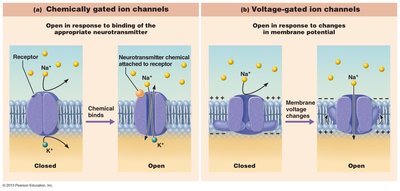
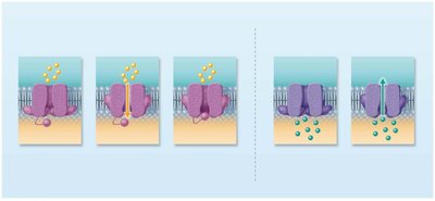
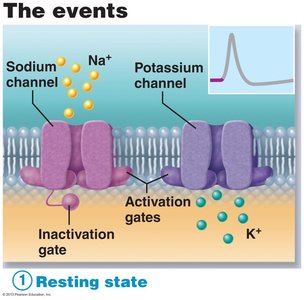
Ion Channels
Leakage Channels: Always open, allow ions to move passively.
Gated Channels: Open/close in response to stimuli; types include chemically gated, voltage-gated, and mechanically gated.
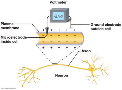
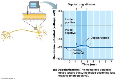
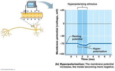
Resting Membrane Potential
Measured at approximately –70 mV in neurons.
Generated by differences in ionic composition and membrane permeability.
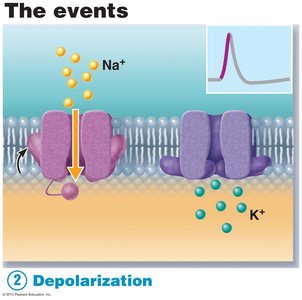
Sodium-potassium pump maintains gradients: 3 Na+ out, 2 K+ in.
Changes in Membrane Potential
Depolarization and Hyperpolarization
Depolarization: Membrane potential becomes less negative (moves toward zero).
Hyperpolarization: Membrane potential becomes more negative (moves away from zero).
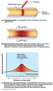
Graded Potentials
Short-lived, localized changes in membrane potential.
Triggered by stimulus opening gated ion channels.
Decay with distance.
Action Potentials
Generation and Propagation
Action potentials are the principal means of long-distance neural communication.
Occur only in muscle cells and axons of neurons.
Do not decay over distance.
Involve opening of voltage-gated channels.
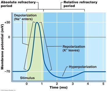
Steps of Action Potential
Resting State: All gated Na+ and K+ channels closed.
Depolarization: Na+ channels open, Na+ influx causes membrane to become more positive.
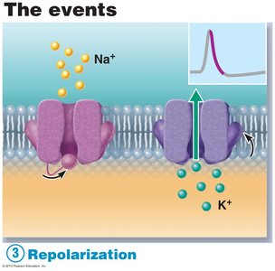
Repolarization: Na+ channels inactivate, K+ channels open, K+ efflux restores negative membrane potential.
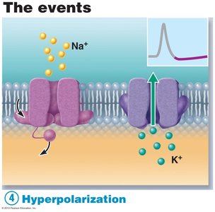
Hyperpolarization: Some K+ channels remain open, membrane potential dips below resting value.
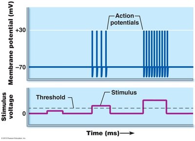
Threshold and All-or-None Principle
AP occurs only if depolarization reaches threshold (~–55 mV).
All-or-None: AP either happens completely or not at all.
Propagation of Action Potential
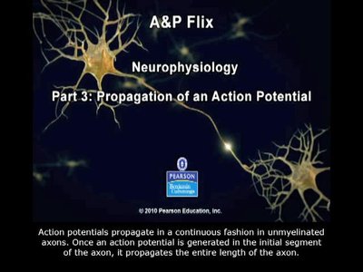
AP is transmitted from origin down entire axon length.
Self-propagating, occurs only in forward direction.
Coding for Stimulus Intensity
All APs are alike; intensity is coded by frequency of impulses.
Higher frequency = stronger stimulus.
Refractory Periods
Absolute Refractory Period: Neuron cannot trigger another AP.
Relative Refractory Period: AP possible only with exceptionally strong stimulus.
The Synapse
Neuron-to-Neuron Communication
Synapses are junctions that mediate information transfer between neurons or from neuron to effector cell.
Presynaptic neuron: Sends information.
Postsynaptic neuron: Receives information.
Chemical Synapses
Most common type; specialized for release and reception of neurotransmitters.
Transmission involves six steps: AP arrival, Ca2+ influx, neurotransmitter release, diffusion and binding, ion channel opening, and termination of neurotransmitter effects.
Electrical Synapses
Less common; neurons are electrically coupled via gap junctions.
Rapid communication, found in some brain regions and embryonic tissue.
Postsynaptic Potentials
Excitatory and Inhibitory Postsynaptic Potentials
EPSP: Depolarizing graded potential, increases likelihood of AP.
IPSP: Hyperpolarizing graded potential, decreases likelihood of AP.
Summation
EPSPs and IPSPs can summate temporally or spatially to influence postsynaptic neuron.
AP is generated only if EPSPs predominate and reach threshold.
Neurotransmitters
Classification by Structure
Acetylcholine (ACh): Released at neuromuscular junctions, degraded by acetylcholinesterase.
Biogenic amines: Includes catecholamines (dopamine, norepinephrine, epinephrine) and indolamines (serotonin, histamine).
Classification by Function
Neurotransmitters can be excitatory or inhibitory, depending on receptor type.
Examples: GABA and glycine (inhibitory), glutamate (excitatory), ACh (excitatory at skeletal muscle, inhibitory at cardiac muscle).
Reflexes
Reflex Arc Components
Reflexes are rapid, automatic responses to stimuli, occurring over pathways called reflex arcs.
Components: receptor, sensory neuron, CNS integration center, motor neuron, effector.
Developmental Aspects of Neurons
Origin of Nervous System
The nervous system originates from the neural tube and neural crest formed from ectoderm. The neural tube becomes the CNS. Additional info: This summary covers the essential concepts of nervous tissue and neurophysiology, including cell types, neuron structure, membrane potentials, synaptic transmission, and neurotransmitter classification, as required for college-level anatomy and physiology courses.
