 Back
BackStudy Guide: Nervous Tissue and the Nervous System
Study Guide - Smart Notes
 Tailored notes based on your materials, expanded with key definitions, examples, and context.
Nervous System Overview
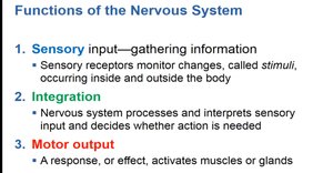
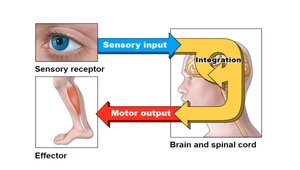
Functions of the Nervous System
The nervous system is responsible for coordinating the body's activities by processing sensory information, integrating it, and producing motor responses.
Sensory input: Gathering information from sensory receptors that monitor changes (stimuli) inside and outside the body.
Integration: Processing and interpreting sensory input to determine if action is needed.
Motor output: Activating muscles or glands in response to processed information.
Nervous Tissue: Neurons
Structure and Function of Neurons
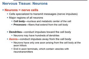
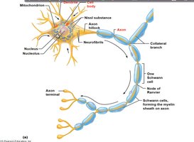
Neurons, or nerve cells, are specialized for transmitting nerve impulses. They consist of distinct regions and processes that facilitate communication within the nervous system.
Cell body: Contains the nucleus and metabolic center.
Processes: Extensions from the cell body, including dendrites and axons.
Dendrites: Conduct impulses toward the cell body; neurons may have many dendrites.
Axons: Conduct impulses away from the cell body; typically only one axon per neuron, ending in terminals with neurotransmitter vesicles.
Synaptic Transmission
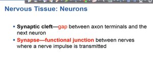
Neurons communicate via synapses, which are functional junctions where nerve impulses are transmitted. The synaptic cleft is the gap between axon terminals and the next neuron.
Synaptic cleft: Gap between axon terminals and the next neuron.
Synapse: Functional junction for impulse transmission.
Myelin Sheath
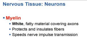
Myelin is a white, fatty material covering axons, providing insulation and increasing the speed of nerve impulse transmission.
Myelin: Protects and insulates fibers; speeds transmission.
Nervous Tissue Terminology
Nervous tissue is classified based on the location and function of its components.
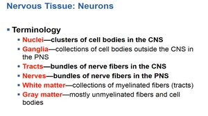
Nuclei: Clusters of cell bodies in the CNS.
Ganglia: Collections of cell bodies outside the CNS in the PNS.
Tracts: Bundles of nerve fibers in the CNS.
Nerves: Bundles of nerve fibers in the PNS.
White matter: Myelinated fibers (tracts).
Gray matter: Unmyelinated fibers and cell bodies.
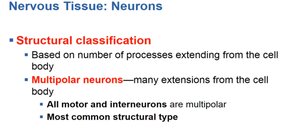
Structural Classification of Neurons
Multipolar and Unipolar Neurons
Neurons are classified by the number of processes extending from the cell body.
Multipolar neurons: Many extensions; all motor and interneurons are multipolar; most common type.
Unipolar neurons: Have a single short process; common in sensory neurons.

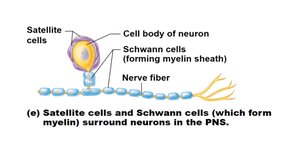
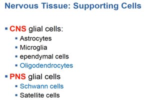
Supporting Cells (Glial Cells)
CNS and PNS Glial Cells
Glial cells support neurons in both the central and peripheral nervous systems.
CNS glial cells: Astrocytes, microglia, ependymal cells, oligodendrocytes.
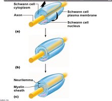
PNS glial cells: Schwann cells, satellite cells.
Central and Peripheral Nervous System
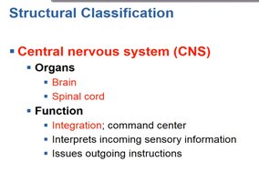
Central Nervous System (CNS)
The CNS consists of the brain and spinal cord, serving as the command center for integration and issuing instructions.
Organs: Brain, spinal cord.
Function: Integration, interpretation of sensory information, issuing instructions.
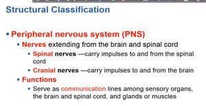
Peripheral Nervous System (PNS)
The PNS includes nerves extending from the CNS, serving as communication lines.
Nerves: Spinal nerves (to/from spinal cord), cranial nerves (to/from brain).
Function: Communication between sensory organs, CNS, glands, and muscles.
Neural Pathways and Sensory Receptors
Neural Pathways
Neural pathways involve afferent (sensory) and efferent (motor) transmission, connecting receptors to effectors via the CNS and PNS.
Afferent transmission: Carries sensory information to the CNS.
Efferent transmission: Carries motor commands from the CNS to effectors.
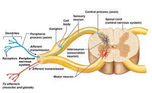
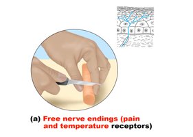
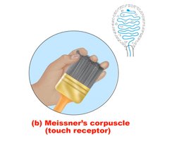
Sensory Receptors
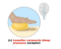
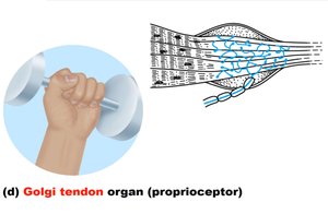
Sensory receptors detect various stimuli, including pain, temperature, touch, pressure, and proprioception.
Free nerve endings: Pain and temperature receptors.
Meissner's corpuscle: Touch receptor.
Lamellar corpuscle: Deep pressure receptor.
Golgi tendon organ: Proprioceptor (detects muscle tension).
Neuronal Membrane Potential and Action Potential
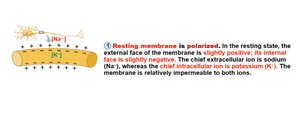
Resting Membrane Potential
The resting membrane is polarized, with the external face slightly positive and the internal face slightly negative.
Chief extracellular ion: Sodium (Na+).
Chief intracellular ion: Potassium (K+).
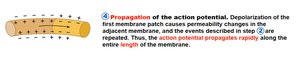
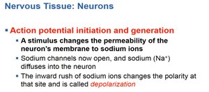
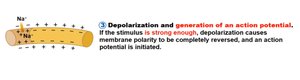
Action Potential Initiation and Generation
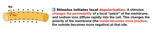
Action potentials are initiated when a stimulus changes the permeability of the neuron's membrane to sodium ions, causing depolarization.
Depolarization: Sodium ions rush in, making the inside more positive.
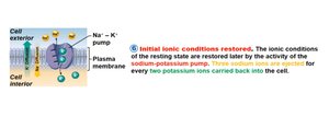
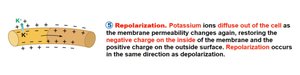
Repolarization and Ionic Restoration
Repolarization occurs as potassium ions diffuse out, restoring the negative charge inside. Ionic conditions are restored by the sodium-potassium pump.
Repolarization: Potassium ions exit, restoring polarity.
Sodium-potassium pump: Restores ionic conditions ().
Summary Table: Nervous Tissue Terminology
Term | Definition |
|---|---|
Nuclei | Clusters of cell bodies in the CNS |
Ganglia | Collections of cell bodies outside the CNS in the PNS |
Tracts | Bundles of nerve fibers in the CNS |
Nerves | Bundles of nerve fibers in the PNS |
White matter | Collections of myelinated fibers (tracts) |
Gray matter | Mostly unmyelinated fibers and cell bodies |
Summary Table: Glial Cells
System | Glial Cell Type | Function |
|---|---|---|
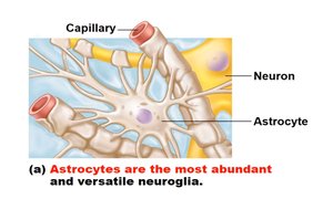
CNS | Astrocytes | Support, nutrient transfer, blood-brain barrier |
CNS | Microglia | Phagocytosis, defense |
CNS | Ependymal cells | Line cerebrospinal fluid-filled cavities |
CNS | Oligodendrocytes | Form myelin sheaths |
PNS | Schwann cells | Form myelin sheaths |
PNS | Satellite cells | Support neuron cell bodies |
Key Equations
Sodium-Potassium Pump
For every three sodium ions () pumped out, two potassium ions () are pumped in:
Membrane Potential
Resting membrane potential is typically:
Additional info:
Expanded explanations of neuron structure, glial cell functions, and action potential steps were added for academic completeness.
